

5.7 Non-Adhesive Motility
107
in the cell corners. In addition to these mechanical effects, adhesion strength and
myosin contraction affect the organization of the actin network through various signal
transduction pathways (recall Fig. 5.16).
5.7 Non-Adhesive Motility
There are different manners of motion, independent of adhesion. The alga Euglena
gracilis, which can also swim by flagellar propulsion, crawls by gracefully reshaping
its whole body (in what is known as metaboly), as shown in Fig. 5.22.
Motion not relying on adhesion and involving frequent shape changes is described
as amoeboid. It is more characteristic of motion in a 3D tissue environment. Its
advantage is the freedom to set routes dictated by the structure of a substrate, and
the ability to rapidly maneuver in a 3D environment following environmental, e.g.,
chemotactic, cues. Leukocytes, scattered throughout the body and able to infiltrate
any type of tissue, follow this strategy, and are able to migrate up to 100 times faster
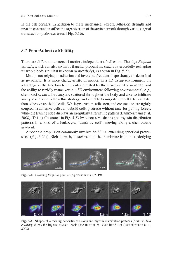
than adhesive epithelial cells. While protrusion, adhesion, and contraction are tightly
coupled in adhesive cells, amoeboid cells protrude without anterior pulling forces,
while the trailing edge displays an irregularly alternating pattern (Lämmermann et al,
2008). This is illustrated in Fig. 5.23 by successive shapes and myosin distribution
patterns in a kind of a leukocyte, “dendritic cell”, moving along a chemotactic
gradient.
Amoeboid propulsion commonly involves blebbing, extending spherical protrusions (Fig. 5.24a). Blebs form by detachment of the membrane from the underlying
Fig. 5.22 Crawling Euglena gracilis (Agostinelli et al, 2019)
Fig. 5.23 Shapes of a moving dendritic cell (top) and myosin distribution patterns (bottom). Red
coloring shows the highest myosin level; time in minutes; scale bar 5 μm (Lämmermann et al,
2008)
107
in the cell corners. In addition to these mechanical effects, adhesion strength and
myosin contraction affect the organization of the actin network through various signal
transduction pathways (recall Fig. 5.16).
5.7 Non-Adhesive Motility
There are different manners of motion, independent of adhesion. The alga Euglena
gracilis, which can also swim by flagellar propulsion, crawls by gracefully reshaping
its whole body (in what is known as metaboly), as shown in Fig. 5.22.
Motion not relying on adhesion and involving frequent shape changes is described
as amoeboid. It is more characteristic of motion in a 3D tissue environment. Its
advantage is the freedom to set routes dictated by the structure of a substrate, and
the ability to rapidly maneuver in a 3D environment following environmental, e.g.,
chemotactic, cues. Leukocytes, scattered throughout the body and able to infiltrate
any type of tissue, follow this strategy, and are able to migrate up to 100 times faster
than adhesive epithelial cells. While protrusion, adhesion, and contraction are tightly
coupled in adhesive cells, amoeboid cells protrude without anterior pulling forces,
while the trailing edge displays an irregularly alternating pattern (Lämmermann et al,
2008). This is illustrated in Fig. 5.23 by successive shapes and myosin distribution
patterns in a kind of a leukocyte, “dendritic cell”, moving along a chemotactic
gradient.
Amoeboid propulsion commonly involves blebbing, extending spherical protrusions (Fig. 5.24a). Blebs form by detachment of the membrane from the underlying
Fig. 5.22 Crawling Euglena gracilis (Agostinelli et al, 2019)
Fig. 5.23 Shapes of a moving dendritic cell (top) and myosin distribution patterns (bottom). Red
coloring shows the highest myosin level; time in minutes; scale bar 5 μm (Lämmermann et al,
2008)