

106
5 Eukaryotic Cells
supported by the observed fluctuations in the leading edge advance velocity in space
(Fig. 5.20b) and time (Fig. 5.20c).
Protrusions may be resisted by tension in the plasma membrane, keeping the leading edge smooth and the shape elongated normally to the direction of motion. This
is characteristic of keratocytes taken from fish skin, favored by experimentalists for
their relatively fast and persistently directed motion. Barnhart et al (2011) revealed
a strong effect of adhesive properties of the underlying substrate on the morphology
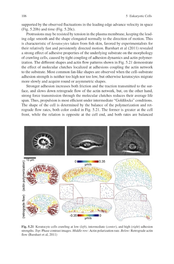
of crawling cells, caused by tight coupling of adhesion dynamics and actin polymerization. The different shapes and actin flow patterns shown in Fig. 5.21 demonstrate
the effect of molecular clutches localized at adhesions coupling the actin network
to the substrate. Most common fan-like shapes are observed when the cell–substrate
adhesion strength is neither too high nor too low, but otherwise keratocytes migrate
more slowly and acquire round or asymmetric shapes.
Stronger adhesion increases both friction and the traction transmitted to the surface, and slows down retrograde flow of the actin network, but, on the other hand,
strong force transmission through the molecular clutches reduces their average life
span. Thus, propulsion is most efficient under intermediate “Goldilocks” conditions.
The shape of the cell is determined by the balance of the polymerization and retrograde flow rates, both color coded in Fig. 5.21. The former is greater at the cell
front, while the relation is opposite at the cell end, and both rates are balanced
Fig. 5.21 Keratocyte cells crawling at low (left), intermediate (center), and high (right) adhesion
strengths. Top: Phase contrast images. Middle row: Actin polarization rate. Below: Retrograde actin
flow (Barnhart et al, 2011)
5 Eukaryotic Cells
supported by the observed fluctuations in the leading edge advance velocity in space
(Fig. 5.20b) and time (Fig. 5.20c).
Protrusions may be resisted by tension in the plasma membrane, keeping the leading edge smooth and the shape elongated normally to the direction of motion. This
is characteristic of keratocytes taken from fish skin, favored by experimentalists for
their relatively fast and persistently directed motion. Barnhart et al (2011) revealed
a strong effect of adhesive properties of the underlying substrate on the morphology
of crawling cells, caused by tight coupling of adhesion dynamics and actin polymerization. The different shapes and actin flow patterns shown in Fig. 5.21 demonstrate
the effect of molecular clutches localized at adhesions coupling the actin network
to the substrate. Most common fan-like shapes are observed when the cell–substrate
adhesion strength is neither too high nor too low, but otherwise keratocytes migrate
more slowly and acquire round or asymmetric shapes.
Stronger adhesion increases both friction and the traction transmitted to the surface, and slows down retrograde flow of the actin network, but, on the other hand,
strong force transmission through the molecular clutches reduces their average life
span. Thus, propulsion is most efficient under intermediate “Goldilocks” conditions.
The shape of the cell is determined by the balance of the polymerization and retrograde flow rates, both color coded in Fig. 5.21. The former is greater at the cell
front, while the relation is opposite at the cell end, and both rates are balanced
Fig. 5.21 Keratocyte cells crawling at low (left), intermediate (center), and high (right) adhesion
strengths. Top: Phase contrast images. Middle row: Actin polarization rate. Below: Retrograde actin
flow (Barnhart et al, 2011)