

5.6 Adhesive Crawling
105
foot, although those are not feet at all but aids to explore the way ahead: they can be
either withdrawn or strengthened by actin filaments to pull the leading edge ahead
(Fig. 5.19a). Nascent adhesions form in the lamellipodium, immediately behind the
leading edge. Their assembly determines the protrusion rate of the leading edge and
requires actin polymerization (see Fig. 5.19b).
Not all nascent adhesions persist, but the persistent ones evolve to larger focal
complexes slightly behind the leading edge, and further into mature focal adhesions
that reside at the ends of large actin bundles or stress fibers that extend to the centre
or the rear of the cell. Maturation involves engagement of the molecular clutch
(Fig. 5.19c), and the resulting traction force on the substrate or extracellular matrix
(ECM) leads to a net edge protrusion.
However, the leading edge is a line rather than a single point, and protrusions
have to be supported by the formation of an actin arc along the leading edge to
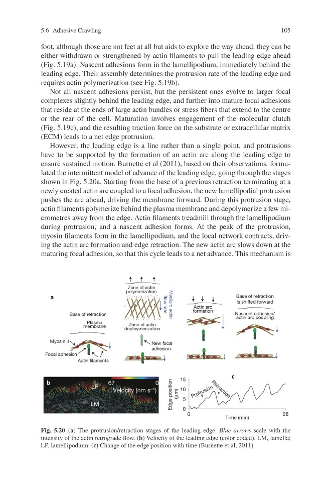
ensure sustained motion. Burnette et al (2011), based on their observations, formulated the intermittent model of advance of the leading edge, going through the stages
shown in Fig. 5.20a. Starting from the base of a previous retraction terminating at a
newly created actin arc coupled to a focal adhesion, the new lamellipodial protrusion
pushes the arc ahead, driving the membrane forward. During this protrusion stage,
actin filaments polymerize behind the plasma membrane and depolymerize a few micrometres away from the edge. Actin filaments treadmill through the lamellipodium
during protrusion, and a nascent adhesion forms. At the peak of the protrusion,
myosin filaments form in the lamellipodium, and the local network contracts, driving the actin arc formation and edge retraction. The new actin arc slows down at the
maturing focal adhesion, so that this cycle leads to a net advance. This mechanism is
Fig. 5.20 (a) The protrusion/retraction stages of the leading edge. Blue arrows scale with the
intensity of the actin retrograde flow. (b) Velocity of the leading edge (color coded). LM, lamella;
LP, lamellipodium. (c) Change of the edge position with time (Burnette et al, 2011)
105
foot, although those are not feet at all but aids to explore the way ahead: they can be
either withdrawn or strengthened by actin filaments to pull the leading edge ahead
(Fig. 5.19a). Nascent adhesions form in the lamellipodium, immediately behind the
leading edge. Their assembly determines the protrusion rate of the leading edge and
requires actin polymerization (see Fig. 5.19b).
Not all nascent adhesions persist, but the persistent ones evolve to larger focal
complexes slightly behind the leading edge, and further into mature focal adhesions
that reside at the ends of large actin bundles or stress fibers that extend to the centre
or the rear of the cell. Maturation involves engagement of the molecular clutch
(Fig. 5.19c), and the resulting traction force on the substrate or extracellular matrix
(ECM) leads to a net edge protrusion.
However, the leading edge is a line rather than a single point, and protrusions
have to be supported by the formation of an actin arc along the leading edge to
ensure sustained motion. Burnette et al (2011), based on their observations, formulated the intermittent model of advance of the leading edge, going through the stages
shown in Fig. 5.20a. Starting from the base of a previous retraction terminating at a
newly created actin arc coupled to a focal adhesion, the new lamellipodial protrusion
pushes the arc ahead, driving the membrane forward. During this protrusion stage,
actin filaments polymerize behind the plasma membrane and depolymerize a few micrometres away from the edge. Actin filaments treadmill through the lamellipodium
during protrusion, and a nascent adhesion forms. At the peak of the protrusion,
myosin filaments form in the lamellipodium, and the local network contracts, driving the actin arc formation and edge retraction. The new actin arc slows down at the
maturing focal adhesion, so that this cycle leads to a net advance. This mechanism is
Fig. 5.20 (a) The protrusion/retraction stages of the leading edge. Blue arrows scale with the
intensity of the actin retrograde flow. (b) Velocity of the leading edge (color coded). LM, lamella;
LP, lamellipodium. (c) Change of the edge position with time (Burnette et al, 2011)