

104
5 Eukaryotic Cells
monomers. The observed transformations among alternative structures are shown in
Fig. 5.18, with prevalent transitions indicated by thick arrows.
5.6 Adhesive Crawling
Unicellular organisms need to move, and isolated cells move toward preferred environments. This is not an easy task when traveling on a solid substrate and having
no flagella or other suitable appendages. Focal adhesions cannot step like feet; they
have to be released at the hind end and created anew ahead, and motion requires
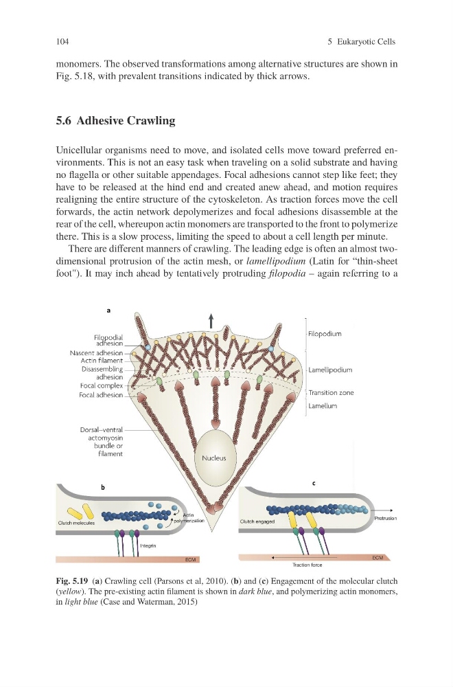
realigning the entire structure of the cytoskeleton. As traction forces move the cell
forwards, the actin network depolymerizes and focal adhesions disassemble at the
rear of the cell, whereupon actin monomers are transported to the front to polymerize
there. This is a slow process, limiting the speed to about a cell length per minute.
There are different manners of crawling. The leading edge is often an almost twodimensional protrusion of the actin mesh, or lamellipodium (Latin for “thin-sheet
foot"). It may inch ahead by tentatively protruding filopodia – again referring to a
Fig. 5.19 (a) Crawling cell (Parsons et al, 2010). (b) and (c) Engagement of the molecular clutch
(yellow). The pre-existing actin filament is shown in dark blue, and polymerizing actin monomers,
in light blue (Case and Waterman, 2015)
5 Eukaryotic Cells
monomers. The observed transformations among alternative structures are shown in
Fig. 5.18, with prevalent transitions indicated by thick arrows.
5.6 Adhesive Crawling
Unicellular organisms need to move, and isolated cells move toward preferred environments. This is not an easy task when traveling on a solid substrate and having
no flagella or other suitable appendages. Focal adhesions cannot step like feet; they
have to be released at the hind end and created anew ahead, and motion requires
realigning the entire structure of the cytoskeleton. As traction forces move the cell
forwards, the actin network depolymerizes and focal adhesions disassemble at the
rear of the cell, whereupon actin monomers are transported to the front to polymerize
there. This is a slow process, limiting the speed to about a cell length per minute.
There are different manners of crawling. The leading edge is often an almost twodimensional protrusion of the actin mesh, or lamellipodium (Latin for “thin-sheet
foot"). It may inch ahead by tentatively protruding filopodia – again referring to a
Fig. 5.19 (a) Crawling cell (Parsons et al, 2010). (b) and (c) Engagement of the molecular clutch
(yellow). The pre-existing actin filament is shown in dark blue, and polymerizing actin monomers,
in light blue (Case and Waterman, 2015)