

5.5 Adhesion
103
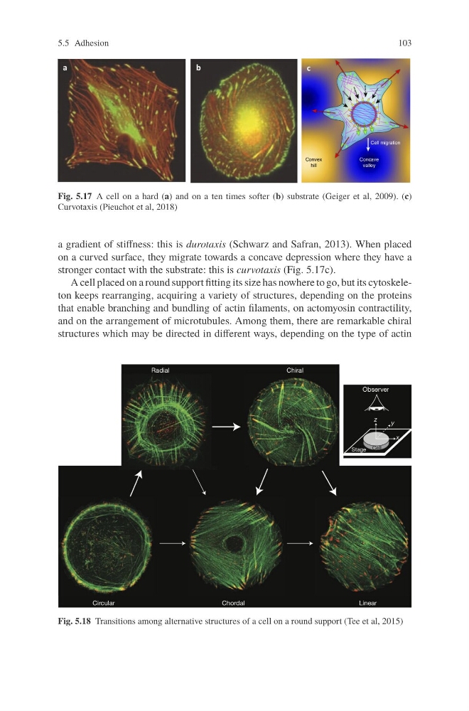
Fig. 5.17 A cell on a hard (a) and on a ten times softer (b) substrate (Geiger et al, 2009). (c)
Curvotaxis (Pieuchot et al, 2018)
a gradient of stiffness: this is durotaxis (Schwarz and Safran, 2013). When placed
on a curved surface, they migrate towards a concave depression where they have a
stronger contact with the substrate: this is curvotaxis (Fig. 5.17c).
A cell placed on a round support fitting its size has nowhere to go, but its cytoskeleton keeps rearranging, acquiring a variety of structures, depending on the proteins
that enable branching and bundling of actin filaments, on actomyosin contractility,
and on the arrangement of microtubules. Among them, there are remarkable chiral
structures which may be directed in different ways, depending on the type of actin
Fig. 5.18 Transitions among alternative structures of a cell on a round support (Tee et al, 2015)
103
Fig. 5.17 A cell on a hard (a) and on a ten times softer (b) substrate (Geiger et al, 2009). (c)
Curvotaxis (Pieuchot et al, 2018)
a gradient of stiffness: this is durotaxis (Schwarz and Safran, 2013). When placed
on a curved surface, they migrate towards a concave depression where they have a
stronger contact with the substrate: this is curvotaxis (Fig. 5.17c).
A cell placed on a round support fitting its size has nowhere to go, but its cytoskeleton keeps rearranging, acquiring a variety of structures, depending on the proteins
that enable branching and bundling of actin filaments, on actomyosin contractility,
and on the arrangement of microtubules. Among them, there are remarkable chiral
structures which may be directed in different ways, depending on the type of actin
Fig. 5.18 Transitions among alternative structures of a cell on a round support (Tee et al, 2015)