

102
5 Eukaryotic Cells
Dorsal SF
Focal adhesions
Transverse arcs Ventral SF
b
c
a
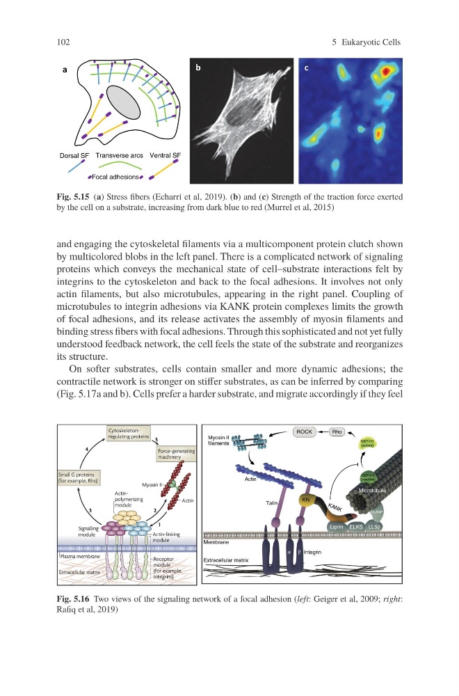
Fig. 5.15 (a) Stress fibers (Echarri et al, 2019). (b) and (c) Strength of the traction force exerted
by the cell on a substrate, increasing from dark blue to red (Murrel et al, 2015)
and engaging the cytoskeletal filaments via a multicomponent protein clutch shown
by multicolored blobs in the left panel. There is a complicated network of signaling
proteins which conveys the mechanical state of cell–substrate interactions felt by
integrins to the cytoskeleton and back to the focal adhesions. It involves not only
actin filaments, but also microtubules, appearing in the right panel. Coupling of
microtubules to integrin adhesions via KANK protein complexes limits the growth
of focal adhesions, and its release activates the assembly of myosin filaments and
binding stress fibers with focal adhesions. Through this sophisticated and not yet fully
understood feedback network, the cell feels the state of the substrate and reorganizes
its structure.
On softer substrates, cells contain smaller and more dynamic adhesions; the
contractile network is stronger on stiffer substrates, as can be inferred by comparing
(Fig. 5.17a and b). Cells prefer a harder substrate, and migrate accordingly if they feel
Fig. 5.16 Two views of the signaling network of a focal adhesion (left: Geiger et al, 2009; right:
Rafiq et al, 2019)
5 Eukaryotic Cells
Dorsal SF
Focal adhesions
Transverse arcs Ventral SF
b
c
a
Fig. 5.15 (a) Stress fibers (Echarri et al, 2019). (b) and (c) Strength of the traction force exerted
by the cell on a substrate, increasing from dark blue to red (Murrel et al, 2015)
and engaging the cytoskeletal filaments via a multicomponent protein clutch shown
by multicolored blobs in the left panel. There is a complicated network of signaling
proteins which conveys the mechanical state of cell–substrate interactions felt by
integrins to the cytoskeleton and back to the focal adhesions. It involves not only
actin filaments, but also microtubules, appearing in the right panel. Coupling of
microtubules to integrin adhesions via KANK protein complexes limits the growth
of focal adhesions, and its release activates the assembly of myosin filaments and
binding stress fibers with focal adhesions. Through this sophisticated and not yet fully
understood feedback network, the cell feels the state of the substrate and reorganizes
its structure.
On softer substrates, cells contain smaller and more dynamic adhesions; the
contractile network is stronger on stiffer substrates, as can be inferred by comparing
(Fig. 5.17a and b). Cells prefer a harder substrate, and migrate accordingly if they feel
Fig. 5.16 Two views of the signaling network of a focal adhesion (left: Geiger et al, 2009; right:
Rafiq et al, 2019)