

5.5 Adhesion
101
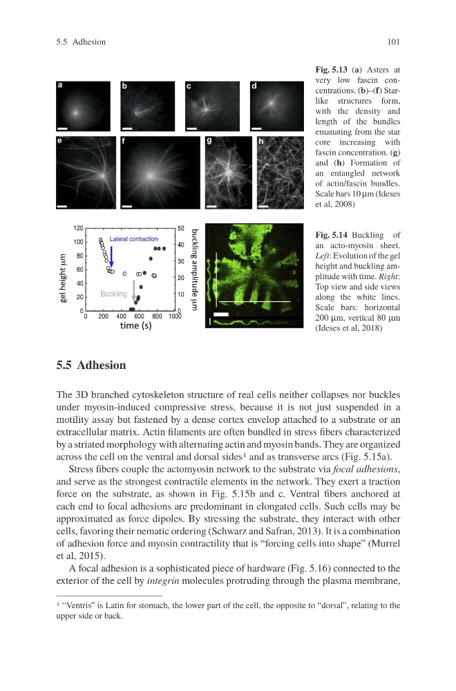
Fig. 5.13 (a) Asters at
very low fascin concentrations. (b)–(f) Starlike structures form,
with the density and
length of the bundles
emanating from the star
core increasing with
fascin concentration. (g)
and (h) Formation of
an entangled network
of actin/fascin bundles.
Scale bars 10 μm (Ideses
et al, 2008)
Fig. 5.14 Buckling of
an acto-myosin sheet.
Left: Evolution of the gel
height and buckling amplitude with time. Right:
Top view and side views
along the white lines.
Scale bars: horizontal
200 μm, vertical 80 μm
(Ideses et al, 2018)
5.5 Adhesion
The 3D branched cytoskeleton structure of real cells neither collapses nor buckles
under myosin-induced compressive stress, because it is not just suspended in a
motility assay but fastened by a dense cortex envelop attached to a substrate or an
extracellular matrix. Actin filaments are often bundled in stress fibers characterized
by a striated morphology with alternating actin and myosin bands. They are organized
across the cell on the ventral and dorsal sides1 and as transverse arcs (Fig. 5.15a).
Stress fibers couple the actomyosin network to the substrate via focal adhesions,
and serve as the strongest contractile elements in the network. They exert a traction
force on the substrate, as shown in Fig. 5.15b and c. Ventral fibers anchored at
each end to focal adhesions are predominant in elongated cells. Such cells may be
approximated as force dipoles. By stressing the substrate, they interact with other
cells, favoring their nematic ordering (Schwarz and Safran, 2013). It is a combination
of adhesion force and myosin contractility that is “forcing cells into shape” (Murrel
et al, 2015).
A focal adhesion is a sophisticated piece of hardware (Fig. 5.16) connected to the
exterior of the cell by integrin molecules protruding through the plasma membrane,
1 “Ventris” is Latin for stomach, the lower part of the cell, the opposite to “dorsal”, relating to the
upper side or back.
101
Fig. 5.13 (a) Asters at
very low fascin concentrations. (b)–(f) Starlike structures form,
with the density and
length of the bundles
emanating from the star
core increasing with
fascin concentration. (g)
and (h) Formation of
an entangled network
of actin/fascin bundles.
Scale bars 10 μm (Ideses
et al, 2008)
Fig. 5.14 Buckling of
an acto-myosin sheet.
Left: Evolution of the gel
height and buckling amplitude with time. Right:
Top view and side views
along the white lines.
Scale bars: horizontal
200 μm, vertical 80 μm
(Ideses et al, 2018)
5.5 Adhesion
The 3D branched cytoskeleton structure of real cells neither collapses nor buckles
under myosin-induced compressive stress, because it is not just suspended in a
motility assay but fastened by a dense cortex envelop attached to a substrate or an
extracellular matrix. Actin filaments are often bundled in stress fibers characterized
by a striated morphology with alternating actin and myosin bands. They are organized
across the cell on the ventral and dorsal sides1 and as transverse arcs (Fig. 5.15a).
Stress fibers couple the actomyosin network to the substrate via focal adhesions,
and serve as the strongest contractile elements in the network. They exert a traction
force on the substrate, as shown in Fig. 5.15b and c. Ventral fibers anchored at
each end to focal adhesions are predominant in elongated cells. Such cells may be
approximated as force dipoles. By stressing the substrate, they interact with other
cells, favoring their nematic ordering (Schwarz and Safran, 2013). It is a combination
of adhesion force and myosin contractility that is “forcing cells into shape” (Murrel
et al, 2015).
A focal adhesion is a sophisticated piece of hardware (Fig. 5.16) connected to the
exterior of the cell by integrin molecules protruding through the plasma membrane,
1 “Ventris” is Latin for stomach, the lower part of the cell, the opposite to “dorsal”, relating to the
upper side or back.