

98
5 Eukaryotic Cells
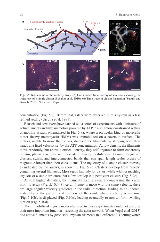
Fig. 5.9 (a) Scheme of the motility array. (b) Color-coded time overlay of snapshots showing the
trajectory of a single cluster (Schaller et al, 2010). (c) Time trace of cluster formation (Suzuki and
Bausch, 2017). Scale bars 50 μm
concentration (Fig. 5.8). Before that, asters were observed in this system in a less
refined setting (Urrutia et al, 1991).
Bausch and coworkers have carried out a series of experiments with a mixture of
actin filaments and myosin motors powered by ATP in a still more constrained setting
of motility assays, schematized in Fig. 5.9a, where a particular kind of molecular
motor (heavy meromyosin HMM) was immobilized on a coverslip surface. The
motors, unable to move themselves, displace the filaments by stepping with their
heads at a fixed velocity set by the ATP concentration. At low density, the filaments
move randomly, but above a critical density, they self-organize to form coherently
moving planar structures with persistent density modulations, forming long-lived
clusters, swirls, and interconnected bands that can span length scales orders of
magnitude longer than their constituents. The trajectory of a single cluster, moving
as indicated by the arrows, is shown in Fig. 5.9b. Clusters develop from “seeds”
containing several filaments. Most seeds last only for a short while without reaching
any sort of a stable structure, but a few develop into persistent clusters (Fig. 5.9c).
At still higher densities, the filaments form a swirl encompassing the entire
motility assay (Fig. 5.10a). Since all filaments move with the same velocity, there
are large angular velocity gradients in the radial direction, leading to an inherent
instability of the pattern, and the core of the swirl, where vorticity is maximal
(Fig. 5.10b), is displaced (Fig. 5.10c), leading eventually to non-uniform swirling
motion (Fig. 5.10d).
The immobilized myosin molecules used in these experiments could not exercise
their most important function – stressing the actin network. When Vogel et al (2013)
tied active filaments by processive myosin filaments in a different 2D setting which
5 Eukaryotic Cells
Fig. 5.9 (a) Scheme of the motility array. (b) Color-coded time overlay of snapshots showing the
trajectory of a single cluster (Schaller et al, 2010). (c) Time trace of cluster formation (Suzuki and
Bausch, 2017). Scale bars 50 μm
concentration (Fig. 5.8). Before that, asters were observed in this system in a less
refined setting (Urrutia et al, 1991).
Bausch and coworkers have carried out a series of experiments with a mixture of
actin filaments and myosin motors powered by ATP in a still more constrained setting
of motility assays, schematized in Fig. 5.9a, where a particular kind of molecular
motor (heavy meromyosin HMM) was immobilized on a coverslip surface. The
motors, unable to move themselves, displace the filaments by stepping with their
heads at a fixed velocity set by the ATP concentration. At low density, the filaments
move randomly, but above a critical density, they self-organize to form coherently
moving planar structures with persistent density modulations, forming long-lived
clusters, swirls, and interconnected bands that can span length scales orders of
magnitude longer than their constituents. The trajectory of a single cluster, moving
as indicated by the arrows, is shown in Fig. 5.9b. Clusters develop from “seeds”
containing several filaments. Most seeds last only for a short while without reaching
any sort of a stable structure, but a few develop into persistent clusters (Fig. 5.9c).
At still higher densities, the filaments form a swirl encompassing the entire
motility assay (Fig. 5.10a). Since all filaments move with the same velocity, there
are large angular velocity gradients in the radial direction, leading to an inherent
instability of the pattern, and the core of the swirl, where vorticity is maximal
(Fig. 5.10b), is displaced (Fig. 5.10c), leading eventually to non-uniform swirling
motion (Fig. 5.10d).
The immobilized myosin molecules used in these experiments could not exercise
their most important function – stressing the actin network. When Vogel et al (2013)
tied active filaments by processive myosin filaments in a different 2D setting which