

5.4 Filaments in Vitro
97
Actin
+TIP
Microtubule
Crosslinker
+
Plasma
membrane
Microtubule
stabilizing
complex
+
+
Fig. 5.7 Interaction between actin filaments and microtubules. Left: Microtubule guidance. Center:
Microtubule anchoring. Right: Actin barrier (Dogterom and Koenderink, 2019)
back when their work is done to use the parts for making other proteins. Internal
signaling and energy exchange play the role of a typesetter. The structure of the
cytoskeleton is the subject of thorough investigation; besides a plethora of papers,
there are quite a few monographs, e.g., Howard (2001), Lee (2013).
5.4 Filaments in Vitro
Away from complexities of the crowded environment of a living cell, biophysicists
study the dynamics of filaments in motility assays containing purified mixtures of
filaments and motors, complete with a supply of ATP sufficient to keep them moving.
Here we step back from live to active matter, where dazzling patterns can be observed,
no longer hidden and unconstrained by realities.
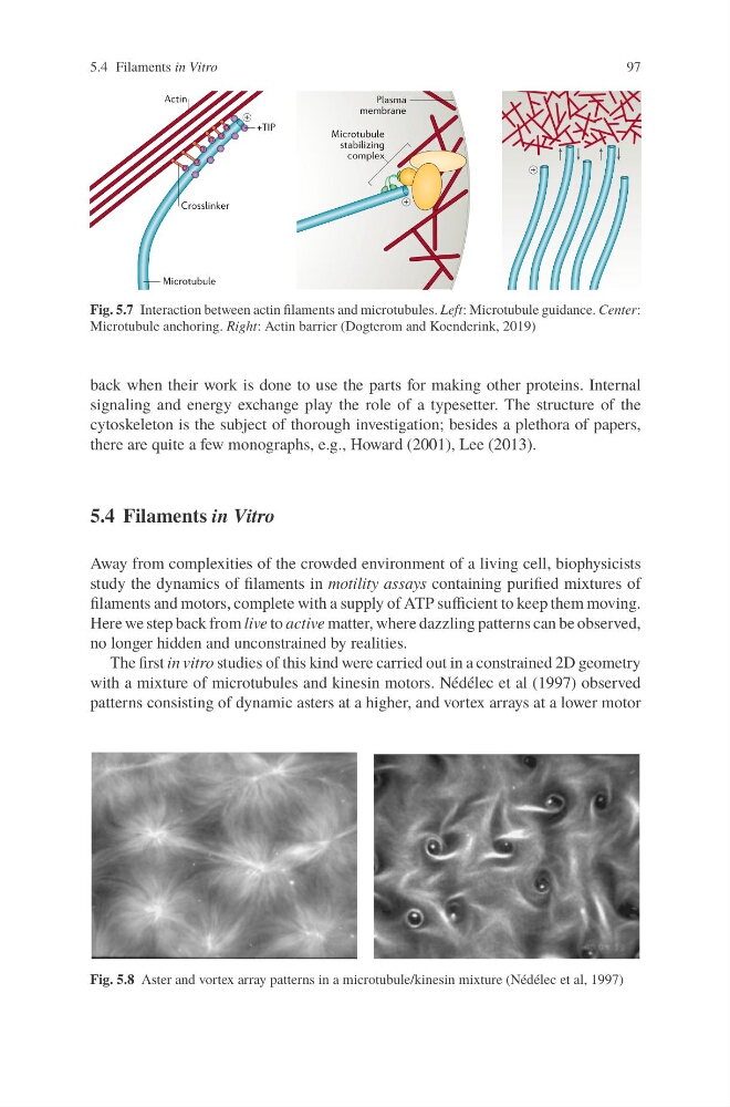
The first in vitro studies of this kind were carried out in a constrained 2D geometry
with a mixture of microtubules and kinesin motors. Nédélec et al (1997) observed
patterns consisting of dynamic asters at a higher, and vortex arrays at a lower motor
Fig. 5.8 Aster and vortex array patterns in a microtubule/kinesin mixture (Nédélec et al, 1997)
97
Actin
+TIP
Microtubule
Crosslinker
+
Plasma
membrane
Microtubule
stabilizing
complex
+
+
Fig. 5.7 Interaction between actin filaments and microtubules. Left: Microtubule guidance. Center:
Microtubule anchoring. Right: Actin barrier (Dogterom and Koenderink, 2019)
back when their work is done to use the parts for making other proteins. Internal
signaling and energy exchange play the role of a typesetter. The structure of the
cytoskeleton is the subject of thorough investigation; besides a plethora of papers,
there are quite a few monographs, e.g., Howard (2001), Lee (2013).
5.4 Filaments in Vitro
Away from complexities of the crowded environment of a living cell, biophysicists
study the dynamics of filaments in motility assays containing purified mixtures of
filaments and motors, complete with a supply of ATP sufficient to keep them moving.
Here we step back from live to active matter, where dazzling patterns can be observed,
no longer hidden and unconstrained by realities.
The first in vitro studies of this kind were carried out in a constrained 2D geometry
with a mixture of microtubules and kinesin motors. Nédélec et al (1997) observed
patterns consisting of dynamic asters at a higher, and vortex arrays at a lower motor
Fig. 5.8 Aster and vortex array patterns in a microtubule/kinesin mixture (Nédélec et al, 1997)