

94
5 Eukaryotic Cells
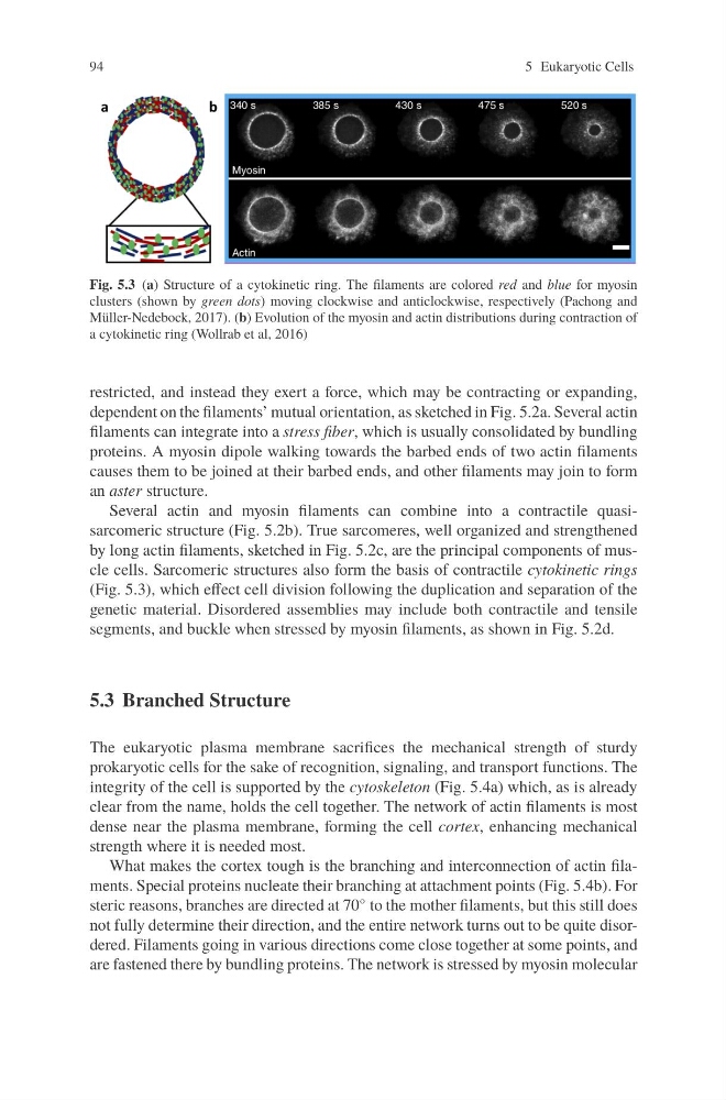
Fig. 5.3 (a) Structure of a cytokinetic ring. The filaments are colored red and blue for myosin
clusters (shown by green dots) moving clockwise and anticlockwise, respectively (Pachong and
Müller-Nedebock, 2017). (b) Evolution of the myosin and actin distributions during contraction of
a cytokinetic ring (Wollrab et al, 2016)
restricted, and instead they exert a force, which may be contracting or expanding,
dependent on the filaments’ mutual orientation, as sketched in Fig. 5.2a. Several actin
filaments can integrate into a stress fiber, which is usually consolidated by bundling
proteins. A myosin dipole walking towards the barbed ends of two actin filaments
causes them to be joined at their barbed ends, and other filaments may join to form
an aster structure.
Several actin and myosin filaments can combine into a contractile quasisarcomeric structure (Fig. 5.2b). True sarcomeres, well organized and strengthened
by long actin filaments, sketched in Fig. 5.2c, are the principal components of muscle cells. Sarcomeric structures also form the basis of contractile cytokinetic rings
(Fig. 5.3), which effect cell division following the duplication and separation of the
genetic material. Disordered assemblies may include both contractile and tensile
segments, and buckle when stressed by myosin filaments, as shown in Fig. 5.2d.
5.3 Branched Structure
The eukaryotic plasma membrane sacrifices the mechanical strength of sturdy
prokaryotic cells for the sake of recognition, signaling, and transport functions. The
integrity of the cell is supported by the cytoskeleton (Fig. 5.4a) which, as is already
clear from the name, holds the cell together. The network of actin filaments is most
dense near the plasma membrane, forming the cell cortex, enhancing mechanical
strength where it is needed most.
What makes the cortex tough is the branching and interconnection of actin filaments. Special proteins nucleate their branching at attachment points (Fig. 5.4b). For
steric reasons, branches are directed at 70 ◦ to the mother filaments, but this still does
not fully determine their direction, and the entire network turns out to be quite disordered. Filaments going in various directions come close together at some points, and
are fastened there by bundling proteins. The network is stressed by myosin molecular
5 Eukaryotic Cells
Fig. 5.3 (a) Structure of a cytokinetic ring. The filaments are colored red and blue for myosin
clusters (shown by green dots) moving clockwise and anticlockwise, respectively (Pachong and
Müller-Nedebock, 2017). (b) Evolution of the myosin and actin distributions during contraction of
a cytokinetic ring (Wollrab et al, 2016)
restricted, and instead they exert a force, which may be contracting or expanding,
dependent on the filaments’ mutual orientation, as sketched in Fig. 5.2a. Several actin
filaments can integrate into a stress fiber, which is usually consolidated by bundling
proteins. A myosin dipole walking towards the barbed ends of two actin filaments
causes them to be joined at their barbed ends, and other filaments may join to form
an aster structure.
Several actin and myosin filaments can combine into a contractile quasisarcomeric structure (Fig. 5.2b). True sarcomeres, well organized and strengthened
by long actin filaments, sketched in Fig. 5.2c, are the principal components of muscle cells. Sarcomeric structures also form the basis of contractile cytokinetic rings
(Fig. 5.3), which effect cell division following the duplication and separation of the
genetic material. Disordered assemblies may include both contractile and tensile
segments, and buckle when stressed by myosin filaments, as shown in Fig. 5.2d.
5.3 Branched Structure
The eukaryotic plasma membrane sacrifices the mechanical strength of sturdy
prokaryotic cells for the sake of recognition, signaling, and transport functions. The
integrity of the cell is supported by the cytoskeleton (Fig. 5.4a) which, as is already
clear from the name, holds the cell together. The network of actin filaments is most
dense near the plasma membrane, forming the cell cortex, enhancing mechanical
strength where it is needed most.
What makes the cortex tough is the branching and interconnection of actin filaments. Special proteins nucleate their branching at attachment points (Fig. 5.4b). For
steric reasons, branches are directed at 70 ◦ to the mother filaments, but this still does
not fully determine their direction, and the entire network turns out to be quite disordered. Filaments going in various directions come close together at some points, and
are fastened there by bundling proteins. The network is stressed by myosin molecular