

Surface Nanostructuring of Metallic Materials …
475
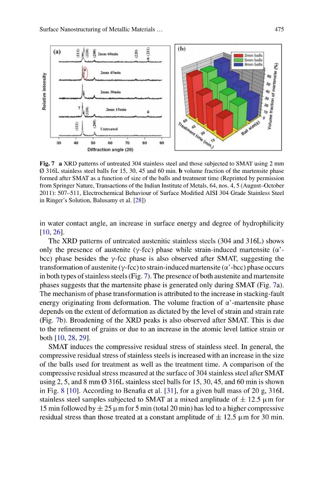
Fig. 7 a XRD patterns of untreated 304 stainless steel and those subjected to SMAT using 2 mm
Ø 316L stainless steel balls for 15, 30, 45 and 60 min. b volume fraction of the martensite phase
formed after SMAT as a function of size of the balls and treatment time (Reprinted by permission
from Springer Nature, Transactions of the Indian Institute of Metals, 64, nos. 4, 5 (August–October
2011): 507–511, Electrochemical Behaviour of Surface Modified AISI 304 Grade Stainless Steel
in Ringer’s Solution, Balusamy et al. [28])
in water contact angle, an increase in surface energy and degree of hydrophilicity
[10, 26].
The XRD patterns of untreated austenitic stainless steels (304 and 316L) shows
only the presence of austenite (γ-fcc) phase while strain-induced martensite (α’bcc) phase besides the γ-fcc phase is also observed after SMAT, suggesting the
transformation of austenite (γ-fcc) to strain-induced martensite (α’-bcc) phase occurs
in both types of stainless steels (Fig. 7). The presence of both austenite and martensite
phases suggests that the martensite phase is generated only during SMAT (Fig. 7a).
The mechanism of phase transformation is attributed to the increase in stacking-fault
energy originating from deformation. The volume fraction of α’-martensite phase
depends on the extent of deformation as dictated by the level of strain and strain rate
(Fig. 7b). Broadening of the XRD peaks is also observed after SMAT. This is due
to the refinement of grains or due to an increase in the atomic level lattice strain or
both [10, 28, 29].
SMAT induces the compressive residual stress of stainless steel. In general, the
compressive residual stress of stainless steels is increased with an increase in the size
of the balls used for treatment as well as the treatment time. A comparison of the
compressive residual stress measured at the surface of 304 stainless steel after SMAT
using 2, 5, and 8 mm Ø 316L stainless steel balls for 15, 30, 45, and 60 min is shown
in Fig. 8 [10]. According to Benafia et al. [31], for a given ball mass of 20 g, 316L
stainless steel samples subjected to SMAT at a mixed amplitude of ± 12.5 μm for
15 min followed by ± 25 μm for 5 min (total 20 min) has led to a higher compressive
residual stress than those treated at a constant amplitude of ± 12.5 μm for 30 min.
475
Fig. 7 a XRD patterns of untreated 304 stainless steel and those subjected to SMAT using 2 mm
Ø 316L stainless steel balls for 15, 30, 45 and 60 min. b volume fraction of the martensite phase
formed after SMAT as a function of size of the balls and treatment time (Reprinted by permission
from Springer Nature, Transactions of the Indian Institute of Metals, 64, nos. 4, 5 (August–October
2011): 507–511, Electrochemical Behaviour of Surface Modified AISI 304 Grade Stainless Steel
in Ringer’s Solution, Balusamy et al. [28])
in water contact angle, an increase in surface energy and degree of hydrophilicity
[10, 26].
The XRD patterns of untreated austenitic stainless steels (304 and 316L) shows
only the presence of austenite (γ-fcc) phase while strain-induced martensite (α’bcc) phase besides the γ-fcc phase is also observed after SMAT, suggesting the
transformation of austenite (γ-fcc) to strain-induced martensite (α’-bcc) phase occurs
in both types of stainless steels (Fig. 7). The presence of both austenite and martensite
phases suggests that the martensite phase is generated only during SMAT (Fig. 7a).
The mechanism of phase transformation is attributed to the increase in stacking-fault
energy originating from deformation. The volume fraction of α’-martensite phase
depends on the extent of deformation as dictated by the level of strain and strain rate
(Fig. 7b). Broadening of the XRD peaks is also observed after SMAT. This is due
to the refinement of grains or due to an increase in the atomic level lattice strain or
both [10, 28, 29].
SMAT induces the compressive residual stress of stainless steel. In general, the
compressive residual stress of stainless steels is increased with an increase in the size
of the balls used for treatment as well as the treatment time. A comparison of the
compressive residual stress measured at the surface of 304 stainless steel after SMAT
using 2, 5, and 8 mm Ø 316L stainless steel balls for 15, 30, 45, and 60 min is shown
in Fig. 8 [10]. According to Benafia et al. [31], for a given ball mass of 20 g, 316L
stainless steel samples subjected to SMAT at a mixed amplitude of ± 12.5 μm for
15 min followed by ± 25 μm for 5 min (total 20 min) has led to a higher compressive
residual stress than those treated at a constant amplitude of ± 12.5 μm for 30 min.