

Nanomaterials: Surface Functionalization …
411
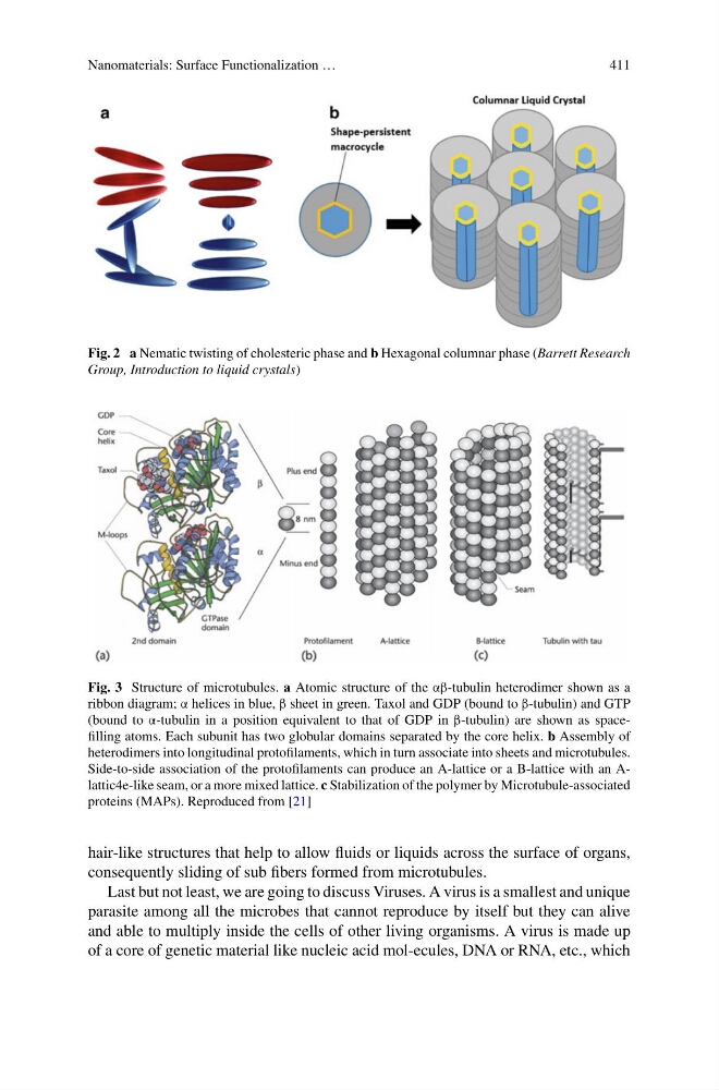
Fig. 2 a Nematic twisting of cholesteric phase and b Hexagonal columnar phase (Barrett Research
Group, Introduction to liquid crystals)
Fig. 3 Structure of microtubules. a Atomic structure of the αβ-tubulin heterodimer shown as a
ribbon diagram; α helices in blue, β sheet in green. Taxol and GDP (bound to β-tubulin) and GTP
(bound to α-tubulin in a position equivalent to that of GDP in β-tubulin) are shown as spacefilling atoms. Each subunit has two globular domains separated by the core helix. b Assembly of
heterodimers into longitudinal protofilaments, which in turn associate into sheets and microtubules.
Side-to-side association of the protofilaments can produce an A-lattice or a B-lattice with an Alattic4e-like seam, or a more mixed lattice. c Stabilization of the polymer by Microtubule-associated
proteins (MAPs). Reproduced from [21]
hair-like structures that help to allow fluids or liquids across the surface of organs,
consequently sliding of sub fibers formed from microtubules.
Last but not least, we are going to discuss Viruses. A virus is a smallest and unique
parasite among all the microbes that cannot reproduce by itself but they can alive
and able to multiply inside the cells of other living organisms. A virus is made up
of a core of genetic material like nucleic acid mol-ecules, DNA or RNA, etc., which
411
Fig. 2 a Nematic twisting of cholesteric phase and b Hexagonal columnar phase (Barrett Research
Group, Introduction to liquid crystals)
Fig. 3 Structure of microtubules. a Atomic structure of the αβ-tubulin heterodimer shown as a
ribbon diagram; α helices in blue, β sheet in green. Taxol and GDP (bound to β-tubulin) and GTP
(bound to α-tubulin in a position equivalent to that of GDP in β-tubulin) are shown as spacefilling atoms. Each subunit has two globular domains separated by the core helix. b Assembly of
heterodimers into longitudinal protofilaments, which in turn associate into sheets and microtubules.
Side-to-side association of the protofilaments can produce an A-lattice or a B-lattice with an Alattic4e-like seam, or a more mixed lattice. c Stabilization of the polymer by Microtubule-associated
proteins (MAPs). Reproduced from [21]
hair-like structures that help to allow fluids or liquids across the surface of organs,
consequently sliding of sub fibers formed from microtubules.
Last but not least, we are going to discuss Viruses. A virus is a smallest and unique
parasite among all the microbes that cannot reproduce by itself but they can alive
and able to multiply inside the cells of other living organisms. A virus is made up
of a core of genetic material like nucleic acid mol-ecules, DNA or RNA, etc., which