

Nanomaterials for Medical Imaging …
359
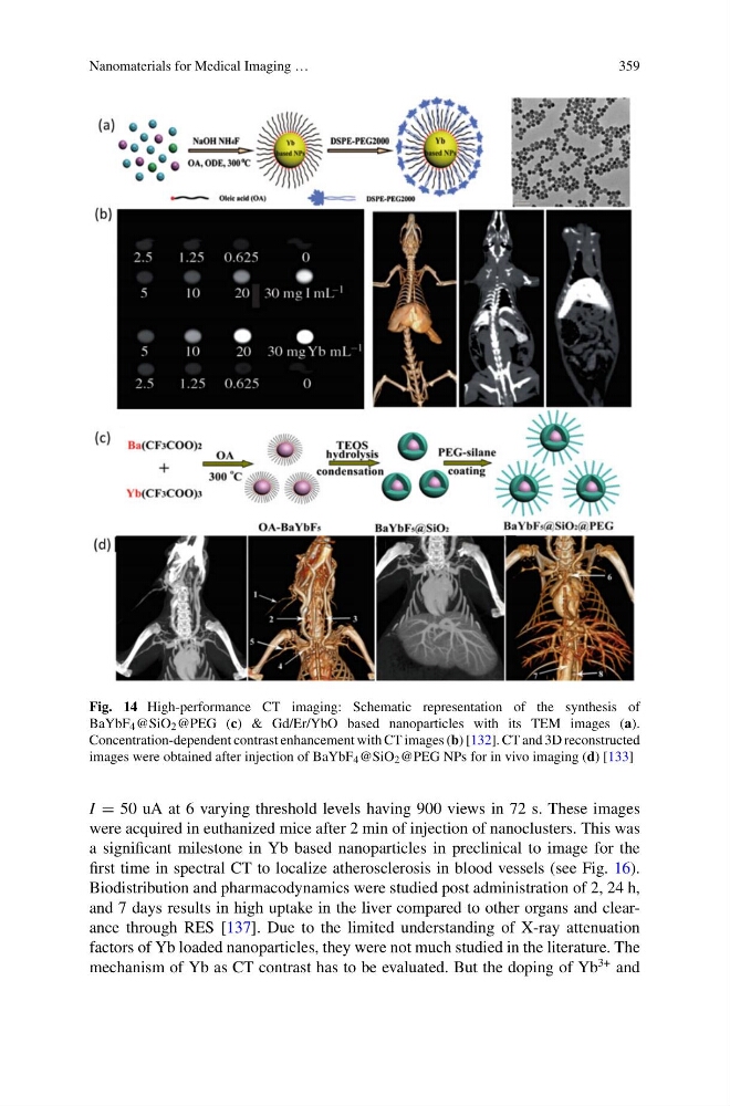
Fig. 14 High-performance CT imaging: Schematic representation of the synthesis of
BaYbF 4 @SiO 2 @PEG (c) & Gd/Er/YbO based nanoparticles with its TEM images (a).
Concentration-dependent contrast enhancement with CT images (b) [132]. CT and 3D reconstructed
images were obtained after injection of BaYbF 4 @SiO 2 @PEG NPs for in vivo imaging (d) [133]
I = 50 uA at 6 varying threshold levels having 900 views in 72 s. These images
were acquired in euthanized mice after 2 min of injection of nanoclusters. This was
a significant milestone in Yb based nanoparticles in preclinical to image for the
first time in spectral CT to localize atherosclerosis in blood vessels (see Fig. 16).
Biodistribution and pharmacodynamics were studied post administration of 2, 24 h,
and 7 days results in high uptake in the liver compared to other organs and clearance through RES [137]. Due to the limited understanding of X-ray attenuation
factors of Yb loaded nanoparticles, they were not much studied in the literature. The
mechanism of Yb as CT contrast has to be evaluated. But the doping of Yb
3+ and
359
Fig. 14 High-performance CT imaging: Schematic representation of the synthesis of
BaYbF 4 @SiO 2 @PEG (c) & Gd/Er/YbO based nanoparticles with its TEM images (a).
Concentration-dependent contrast enhancement with CT images (b) [132]. CT and 3D reconstructed
images were obtained after injection of BaYbF 4 @SiO 2 @PEG NPs for in vivo imaging (d) [133]
I = 50 uA at 6 varying threshold levels having 900 views in 72 s. These images
were acquired in euthanized mice after 2 min of injection of nanoclusters. This was
a significant milestone in Yb based nanoparticles in preclinical to image for the
first time in spectral CT to localize atherosclerosis in blood vessels (see Fig. 16).
Biodistribution and pharmacodynamics were studied post administration of 2, 24 h,
and 7 days results in high uptake in the liver compared to other organs and clearance through RES [137]. Due to the limited understanding of X-ray attenuation
factors of Yb loaded nanoparticles, they were not much studied in the literature. The
mechanism of Yb as CT contrast has to be evaluated. But the doping of Yb
3+ and