

328
G. Perumal and M. Doble
architecture with three-dimensional orientation. These factors are highly important
for enhanced cell functions in vitro and in vivo.
The biomaterials employed in electrospinning were naturally occurring macromolecules, namely collagen, chitosan, silk fibroin, and biodegradable polymers
synthesized via chemical routes such as PGA, PLGA, PLLA, and PCL and blends of
both polymers. Furthermore, several constituents, such as proteins, growth factors,
and hydroxyapatite, can also be combined and developed as nanofibrous materials.
Based on these nano-features, the electrospun scaffolds are capable of supporting
cell adhesion, proliferation, and differentiation in a three-dimensional (3D) manner.
Additionally, the cells grown on these structures lead to biomimetic support for
enhanced cellular functions [27]. However, considerable limitations persist with
this technique for developing intricate 3D scaffold structures, and porous structures
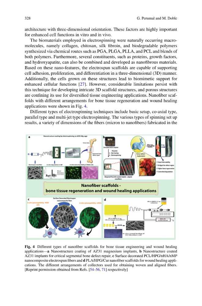
are confining its use for diversified tissue engineering applications. Nanofiber scaffolds with different arrangements for bone tissue regeneration and wound healing
applications were shown in Fig. 4.
Different types of electrospinning techniques include basic setup, co-axial type,
parallel type and multi-jet type electrospinning. The various types of spinning set up
results, a variety of dimensions of the fibers (micron to nanofibers) fabricated in the
Fig. 4 Different types of nanofiber scaffolds for bone tissue engineering and wound healing
applications—a Nanostructure coating of AZ31 magnesium implants, b Nanostructure coated
AZ31 implants for critical segmental bone defect repair, c Surface decorated PCL/HPG/nHA/nMP
nanocomposite electrospun fibers and d PLA/HPG/Cur nanofiber scaffolds for wound healing applications. The different arrangements of collectors used for obtaining woven and aligned fibers.
[Reprint permission obtained from Refs. [54–56, 71] respectively]
G. Perumal and M. Doble
architecture with three-dimensional orientation. These factors are highly important
for enhanced cell functions in vitro and in vivo.
The biomaterials employed in electrospinning were naturally occurring macromolecules, namely collagen, chitosan, silk fibroin, and biodegradable polymers
synthesized via chemical routes such as PGA, PLGA, PLLA, and PCL and blends of
both polymers. Furthermore, several constituents, such as proteins, growth factors,
and hydroxyapatite, can also be combined and developed as nanofibrous materials.
Based on these nano-features, the electrospun scaffolds are capable of supporting
cell adhesion, proliferation, and differentiation in a three-dimensional (3D) manner.
Additionally, the cells grown on these structures lead to biomimetic support for
enhanced cellular functions [27]. However, considerable limitations persist with
this technique for developing intricate 3D scaffold structures, and porous structures
are confining its use for diversified tissue engineering applications. Nanofiber scaffolds with different arrangements for bone tissue regeneration and wound healing
applications were shown in Fig. 4.
Different types of electrospinning techniques include basic setup, co-axial type,
parallel type and multi-jet type electrospinning. The various types of spinning set up
results, a variety of dimensions of the fibers (micron to nanofibers) fabricated in the
Fig. 4 Different types of nanofiber scaffolds for bone tissue engineering and wound healing
applications—a Nanostructure coating of AZ31 magnesium implants, b Nanostructure coated
AZ31 implants for critical segmental bone defect repair, c Surface decorated PCL/HPG/nHA/nMP
nanocomposite electrospun fibers and d PLA/HPG/Cur nanofiber scaffolds for wound healing applications. The different arrangements of collectors used for obtaining woven and aligned fibers.
[Reprint permission obtained from Refs. [54–56, 71] respectively]