

478
M. Rashid et al.
This property of nuclei not to resonate at same frequency arises because of variable electronic environment of nuclei surrounded by particular number of valence
electrons or variable electronic density. This property decides ‘position of signals’
of protons of organic molecules in
1 H-NMR spectrum and hence their nature i.e.
adjacent to electron donating or releasing groups, aromatic, aliphatic etc. [6].
The relative energy of resonance of a particular nucleus resulting from its local environment
is called chemical shift.
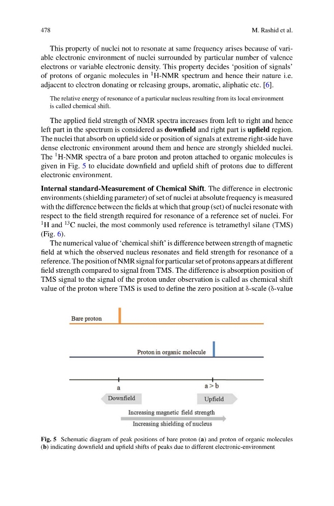
The applied field strength of NMR spectra increases from left to right and hence
left part in the spectrum is considered as downfield and right part is upfield region.
The nuclei that absorb on upfield side or position of signals at extreme right-side have
dense electronic environment around them and hence are strongly shielded nuclei.
The
1 H-NMR spectra of a bare proton and proton attached to organic molecules is
given in Fig. 5 to elucidate downfield and upfield shift of protons due to different
electronic environment.
Internal standard-Measurement of Chemical Shift. The difference in electronic
environments (shielding parameter) of set of nuclei at absolute frequency is measured
with the difference between the fields at which that group (set) of nuclei resonate with
respect to the field strength required for resonance of a reference set of nuclei. For
1 H and
13 C nuclei, the most commonly used reference is tetramethyl silane (TMS)
(Fig. 6).
The numerical value of ‘chemical shift’ is difference between strength of magnetic
field at which the observed nucleus resonates and field strength for resonance of a
reference. The position of NMR signal for particular set of protons appears at different
field strength compared to signal from TMS. The difference is absorption position of
TMS signal to the signal of the proton under observation is called as chemical shift
value of the proton where TMS is used to define the zero position at δ-scale (δ-value
Fig. 5 Schematic diagram of peak positions of bare proton (a) and proton of organic molecules
(b) indicating downfield and upfield shifts of peaks due to different electronic-environment
M. Rashid et al.
This property of nuclei not to resonate at same frequency arises because of variable electronic environment of nuclei surrounded by particular number of valence
electrons or variable electronic density. This property decides ‘position of signals’
of protons of organic molecules in
1 H-NMR spectrum and hence their nature i.e.
adjacent to electron donating or releasing groups, aromatic, aliphatic etc. [6].
The relative energy of resonance of a particular nucleus resulting from its local environment
is called chemical shift.
The applied field strength of NMR spectra increases from left to right and hence
left part in the spectrum is considered as downfield and right part is upfield region.
The nuclei that absorb on upfield side or position of signals at extreme right-side have
dense electronic environment around them and hence are strongly shielded nuclei.
The
1 H-NMR spectra of a bare proton and proton attached to organic molecules is
given in Fig. 5 to elucidate downfield and upfield shift of protons due to different
electronic environment.
Internal standard-Measurement of Chemical Shift. The difference in electronic
environments (shielding parameter) of set of nuclei at absolute frequency is measured
with the difference between the fields at which that group (set) of nuclei resonate with
respect to the field strength required for resonance of a reference set of nuclei. For
1 H and
13 C nuclei, the most commonly used reference is tetramethyl silane (TMS)
(Fig. 6).
The numerical value of ‘chemical shift’ is difference between strength of magnetic
field at which the observed nucleus resonates and field strength for resonance of a
reference. The position of NMR signal for particular set of protons appears at different
field strength compared to signal from TMS. The difference is absorption position of
TMS signal to the signal of the proton under observation is called as chemical shift
value of the proton where TMS is used to define the zero position at δ-scale (δ-value
Fig. 5 Schematic diagram of peak positions of bare proton (a) and proton of organic molecules
(b) indicating downfield and upfield shifts of peaks due to different electronic-environment