

228
T. Umakoshi and P. Verma
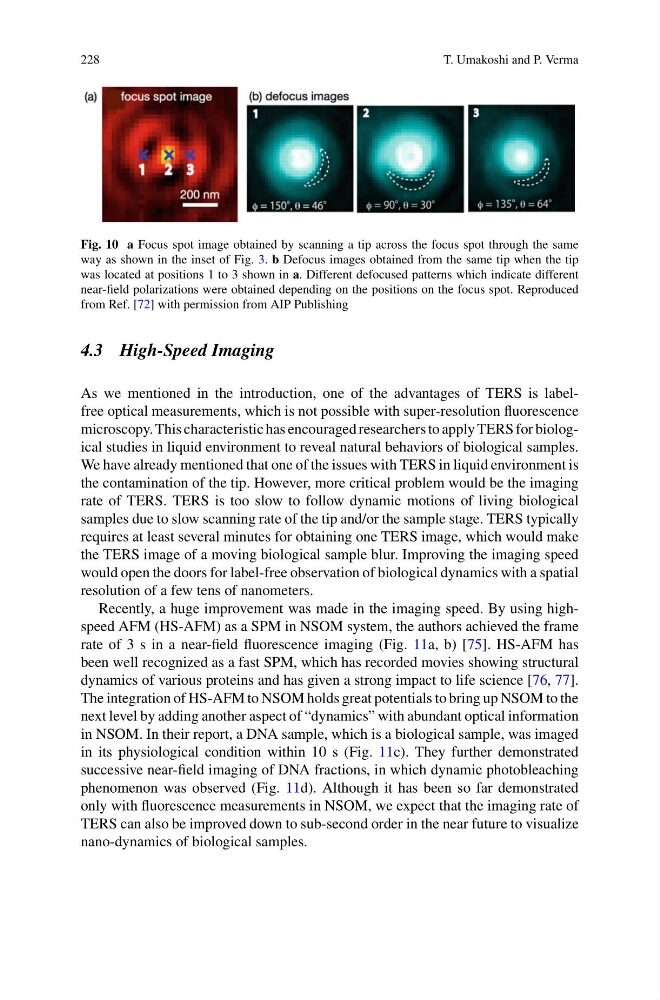
Fig. 10 a Focus spot image obtained by scanning a tip across the focus spot through the same
way as shown in the inset of Fig. 3. b Defocus images obtained from the same tip when the tip
was located at positions 1 to 3 shown in a. Different defocused patterns which indicate different
near-field polarizations were obtained depending on the positions on the focus spot. Reproduced
from Ref. [72] with permission from AIP Publishing
4.3 High-Speed Imaging
As we mentioned in the introduction, one of the advantages of TERS is labelfree optical measurements, which is not possible with super-resolution fluorescence
microscopy. This characteristic has encouraged researchers to apply TERS for biological studies in liquid environment to reveal natural behaviors of biological samples.
We have already mentioned that one of the issues with TERS in liquid environment is
the contamination of the tip. However, more critical problem would be the imaging
rate of TERS. TERS is too slow to follow dynamic motions of living biological
samples due to slow scanning rate of the tip and/or the sample stage. TERS typically
requires at least several minutes for obtaining one TERS image, which would make
the TERS image of a moving biological sample blur. Improving the imaging speed
would open the doors for label-free observation of biological dynamics with a spatial
resolution of a few tens of nanometers.
Recently, a huge improvement was made in the imaging speed. By using highspeed AFM (HS-AFM) as a SPM in NSOM system, the authors achieved the frame
rate of 3 s in a near-field fluorescence imaging (Fig. 11a, b) [75]. HS-AFM has
been well recognized as a fast SPM, which has recorded movies showing structural
dynamics of various proteins and has given a strong impact to life science [76, 77].
The integration of HS-AFM to NSOM holds great potentials to bring up NSOM to the
next level by adding another aspect of “dynamics” with abundant optical information
in NSOM. In their report, a DNA sample, which is a biological sample, was imaged
in its physiological condition within 10 s (Fig. 11c). They further demonstrated
successive near-field imaging of DNA fractions, in which dynamic photobleaching
phenomenon was observed (Fig. 11d). Although it has been so far demonstrated
only with fluorescence measurements in NSOM, we expect that the imaging rate of
TERS can also be improved down to sub-second order in the near future to visualize
nano-dynamics of biological samples.
T. Umakoshi and P. Verma
Fig. 10 a Focus spot image obtained by scanning a tip across the focus spot through the same
way as shown in the inset of Fig. 3. b Defocus images obtained from the same tip when the tip
was located at positions 1 to 3 shown in a. Different defocused patterns which indicate different
near-field polarizations were obtained depending on the positions on the focus spot. Reproduced
from Ref. [72] with permission from AIP Publishing
4.3 High-Speed Imaging
As we mentioned in the introduction, one of the advantages of TERS is labelfree optical measurements, which is not possible with super-resolution fluorescence
microscopy. This characteristic has encouraged researchers to apply TERS for biological studies in liquid environment to reveal natural behaviors of biological samples.
We have already mentioned that one of the issues with TERS in liquid environment is
the contamination of the tip. However, more critical problem would be the imaging
rate of TERS. TERS is too slow to follow dynamic motions of living biological
samples due to slow scanning rate of the tip and/or the sample stage. TERS typically
requires at least several minutes for obtaining one TERS image, which would make
the TERS image of a moving biological sample blur. Improving the imaging speed
would open the doors for label-free observation of biological dynamics with a spatial
resolution of a few tens of nanometers.
Recently, a huge improvement was made in the imaging speed. By using highspeed AFM (HS-AFM) as a SPM in NSOM system, the authors achieved the frame
rate of 3 s in a near-field fluorescence imaging (Fig. 11a, b) [75]. HS-AFM has
been well recognized as a fast SPM, which has recorded movies showing structural
dynamics of various proteins and has given a strong impact to life science [76, 77].
The integration of HS-AFM to NSOM holds great potentials to bring up NSOM to the
next level by adding another aspect of “dynamics” with abundant optical information
in NSOM. In their report, a DNA sample, which is a biological sample, was imaged
in its physiological condition within 10 s (Fig. 11c). They further demonstrated
successive near-field imaging of DNA fractions, in which dynamic photobleaching
phenomenon was observed (Fig. 11d). Although it has been so far demonstrated
only with fluorescence measurements in NSOM, we expect that the imaging rate of
TERS can also be improved down to sub-second order in the near future to visualize
nano-dynamics of biological samples.