

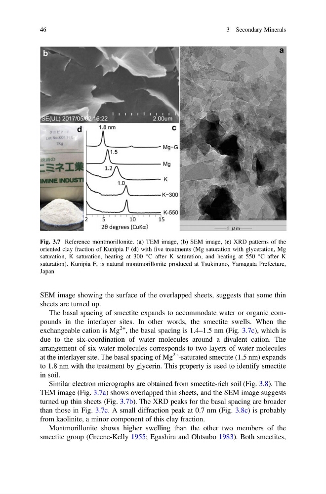
SEM image showing the surface of the overlapped sheets, suggests that some thin
sheets are turned up.
The basal spacing of smectite expands to accommodate water or organic compounds in the interlayer sites. In other words, the smectite swells. When the
exchangeable cation is Mg
2+ , the basal spacing is 1.4–1.5 nm (Fig. 3.7c), which is
due to the six-coordination of water molecules around a divalent cation. The
arrangement of six water molecules corresponds to two layers of water molecules
at the interlayer site. The basal spacing of Mg
2+ -saturated smectite (1.5 nm) expands
to 1.8 nm with the treatment by glycerin. This property is used to identify smectite
in soil.
Similar electron micrographs are obtained from smectite-rich soil (Fig. 3.8). The
TEM image (Fig. 3.7a) shows overlapped thin sheets, and the SEM image suggests
turned up thin sheets (Fig. 3.7b). The XRD peaks for the basal spacing are broader
than those in Fig. 3.7c. A small diffraction peak at 0.7 nm (Fig. 3.8c) is probably
from kaolinite, a minor component of this clay fraction.
Montmorillonite shows higher swelling than the other two members of the
smectite group (Greene-Kelly 1955; Egashira and Ohtsubo 1983). Both smectites,
Fig. 3.7 Reference montmorillonite. (a) TEM image, (b) SEM image, (c) XRD patterns of the
oriented clay fraction of Kunipia F (d) with five treatments (Mg saturation with glyceration, Mg
saturation, K saturation, heating at 300
C after K saturation, and heating at 550
C after K
saturation). Kunipia F, is natural montmorillonite produced at Tsukinuno, Yamagata Prefecture,
Japan
46
3 Secondary Minerals
sheets are turned up.
The basal spacing of smectite expands to accommodate water or organic compounds in the interlayer sites. In other words, the smectite swells. When the
exchangeable cation is Mg
2+ , the basal spacing is 1.4–1.5 nm (Fig. 3.7c), which is
due to the six-coordination of water molecules around a divalent cation. The
arrangement of six water molecules corresponds to two layers of water molecules
at the interlayer site. The basal spacing of Mg
2+ -saturated smectite (1.5 nm) expands
to 1.8 nm with the treatment by glycerin. This property is used to identify smectite
in soil.
Similar electron micrographs are obtained from smectite-rich soil (Fig. 3.8). The
TEM image (Fig. 3.7a) shows overlapped thin sheets, and the SEM image suggests
turned up thin sheets (Fig. 3.7b). The XRD peaks for the basal spacing are broader
than those in Fig. 3.7c. A small diffraction peak at 0.7 nm (Fig. 3.8c) is probably
from kaolinite, a minor component of this clay fraction.
Montmorillonite shows higher swelling than the other two members of the
smectite group (Greene-Kelly 1955; Egashira and Ohtsubo 1983). Both smectites,
Fig. 3.7 Reference montmorillonite. (a) TEM image, (b) SEM image, (c) XRD patterns of the
oriented clay fraction of Kunipia F (d) with five treatments (Mg saturation with glyceration, Mg
saturation, K saturation, heating at 300
C after K saturation, and heating at 550
C after K
saturation). Kunipia F, is natural montmorillonite produced at Tsukinuno, Yamagata Prefecture,
Japan
46
3 Secondary Minerals