

DETERMINATION OF NUCLEOTIDE SEQUENCE
565
O
O
CH 2
N
N
N
N
O
adenine
NH 2
O
N
N
O
NH 2
O CH 2
O
cytosine
O
N
NH
O
O
H 3 C
O CH 2
O
thymine
H 2 N NH 2
H 2 N NH 2
3
6
O
O
CH 2
N
N
N
N
O
NH 2
Me
Me 2 SO 4
OH
ring cleavage
ring cleavage
ring cleavage
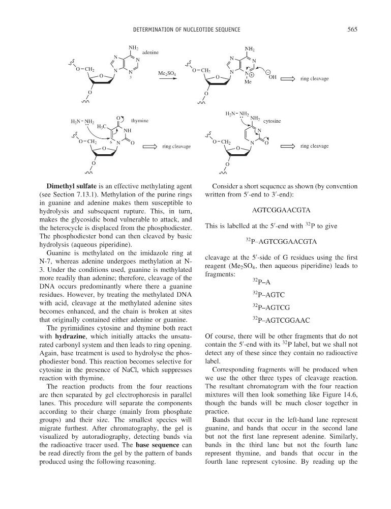
Dimethyl sulfate is an effective methylating agent
(see Section 7.13.1). Methylation of the purine rings
in guanine and adenine makes them susceptible to
hydrolysis and subsequent rupture. This, in turn,
makes the glycosidic bond vulnerable to attack, and
the heterocycle is displaced from the phosphodiester.
The phosphodiester bond can then cleaved by basic
hydrolysis (aqueous piperidine).
Guanine is methylated on the imidazole ring at
N-7, whereas adenine undergoes methylation at N3. Under the conditions used, guanine is methylated
more readily than adenine; therefore, cleavage of the
DNA occurs predominantly where there a guanine
residues. However, by treating the methylated DNA
with acid, cleavage at the methylated adenine sites
becomes enhanced, and the chain is broken at sites
that originally contained either adenine or guanine.
The pyrimidines cytosine and thymine both react
with hydrazine, which initially attacks the unsaturated carbonyl system and then leads to ring opening.
Again, base treatment is used to hydrolyse the phosphodiester bond. This reaction becomes selective for
cytosine in the presence of NaCl, which suppresses
reaction with thymine.
The reaction products from the four reactions
are then separated by gel electrophoresis in parallel
lanes. This procedure will separate the components
according to their charge (mainly from phosphate
groups) and their size. The smallest species will
migrate furthest. After chromatography, the gel is
visualized by autoradiography, detecting bands via
the radioactive tracer used. The base sequence can
be read directly from the gel by the pattern of bands
produced using the following reasoning.
Consider a short sequence as shown (by convention
written from 5
-end to 3
-end):
AGTCGGAACGTA
This is labelled at the 5
-end with
32 P to give
32 P–AGTCGGAACGTA
cleavage at the 5
-side of G residues using the first
reagent (Me 2 SO 4 , then aqueous piperidine) leads to
fragments:
32 P–A
32 P–AGTC
32 P–AGTCG
32 P–AGTCGGAAC
Of course, there will be other fragments that do not
contain the 5
-end with its
32 P label, but we shall not
detect any of these since they contain no radioactive
label.
Corresponding fragments will be produced when
we use the other three types of cleavage reaction.
The resultant chromatogram with the four reaction
mixtures will then look something like Figure 14.6,
though the bands will be much closer together in
practice.
Bands that occur in the left-hand lane represent
guanine, and bands that occur in the second lane
but not the first lane represent adenine. Similarly,
bands in the third lane but not the fourth lane
represent thymine, and bands that occur in the
fourth lane represent cytosine. By reading up the
565
O
O
CH 2
N
N
N
N
O
adenine
NH 2
O
N
N
O
NH 2
O CH 2
O
cytosine
O
N
NH
O
O
H 3 C
O CH 2
O
thymine
H 2 N NH 2
H 2 N NH 2
3
6
O
O
CH 2
N
N
N
N
O
NH 2
Me
Me 2 SO 4
OH
ring cleavage
ring cleavage
ring cleavage
Dimethyl sulfate is an effective methylating agent
(see Section 7.13.1). Methylation of the purine rings
in guanine and adenine makes them susceptible to
hydrolysis and subsequent rupture. This, in turn,
makes the glycosidic bond vulnerable to attack, and
the heterocycle is displaced from the phosphodiester.
The phosphodiester bond can then cleaved by basic
hydrolysis (aqueous piperidine).
Guanine is methylated on the imidazole ring at
N-7, whereas adenine undergoes methylation at N3. Under the conditions used, guanine is methylated
more readily than adenine; therefore, cleavage of the
DNA occurs predominantly where there a guanine
residues. However, by treating the methylated DNA
with acid, cleavage at the methylated adenine sites
becomes enhanced, and the chain is broken at sites
that originally contained either adenine or guanine.
The pyrimidines cytosine and thymine both react
with hydrazine, which initially attacks the unsaturated carbonyl system and then leads to ring opening.
Again, base treatment is used to hydrolyse the phosphodiester bond. This reaction becomes selective for
cytosine in the presence of NaCl, which suppresses
reaction with thymine.
The reaction products from the four reactions
are then separated by gel electrophoresis in parallel
lanes. This procedure will separate the components
according to their charge (mainly from phosphate
groups) and their size. The smallest species will
migrate furthest. After chromatography, the gel is
visualized by autoradiography, detecting bands via
the radioactive tracer used. The base sequence can
be read directly from the gel by the pattern of bands
produced using the following reasoning.
Consider a short sequence as shown (by convention
written from 5
-end to 3
-end):
AGTCGGAACGTA
This is labelled at the 5
-end with
32 P to give
32 P–AGTCGGAACGTA
cleavage at the 5
-side of G residues using the first
reagent (Me 2 SO 4 , then aqueous piperidine) leads to
fragments:
32 P–A
32 P–AGTC
32 P–AGTCG
32 P–AGTCGGAAC
Of course, there will be other fragments that do not
contain the 5
-end with its
32 P label, but we shall not
detect any of these since they contain no radioactive
label.
Corresponding fragments will be produced when
we use the other three types of cleavage reaction.
The resultant chromatogram with the four reaction
mixtures will then look something like Figure 14.6,
though the bands will be much closer together in
practice.
Bands that occur in the left-hand lane represent
guanine, and bands that occur in the second lane
but not the first lane represent adenine. Similarly,
bands in the third lane but not the fourth lane
represent thymine, and bands that occur in the
fourth lane represent cytosine. By reading up the