

NUCLEIC ACIDS
555
of diphosphate to two molecules of phosphate provides some of the driving force to facilitate the reaction (compare Box 14.3 and Section 15.4.1).
14.2.3 RNA
RNA differs structurally from DNA in three important ways. First, as indicated above, the sugar in
RNA is ribose, not 2-deoxyribose. Second, thymine
is replaced by uracil, so that the four bases are adenine, uracil, guanine, and cytosine. The third difference is that RNA is usually single stranded. Although
an RNA molecule may be single stranded, it does
not exclude the possibility of partial double-stranded
sequences being present. In such cases, the molecule
doubles back on itself, and coils up with a complementary base sequence elsewhere. Remember that
complementary sequences now involve A–U rather
than A–T hydrogen-bonding interactions.
DNA stores the genetic information for a cell,
but it is RNA that participates in the processes
by which this information is used. RNA molecules
are classified according to their function or cellular
location. Three major forms are found in prokaryotic
cells:
• messenger RNA (mRNA) carries genetic information from DNA to ribosomes, the organelles
responsible for protein synthesis;
• ribosomal RNA (rRNA) is an integral part of the
ribosomes;
• transfer RNA (tRNA) carries the amino acid
residues that are added to the growing peptide
chain during protein synthesis.
14.2.4 The genetic code
It is the sequence of bases along one of the
strands of the DNA molecule, the coding strand,
that provides the information for the synthesis of
proteins, especially enzymes, in an organism. A
complementary sequence exists along the second
strand, and this is termed the template strand.
A gene is that segment of DNA that contains
the information necessary for the synthesis of one
protein.
Each amino acid in a protein is specified by a
sequence of three nucleotides, termed a codon. A
codon is usually designated in terms of the base
sequence, however, just as we saw with nucleic
acid sequences above. With four different bases,
there are 4
3
= 64 different combinations of three
bases (codons) available, more than enough for the
20 different amino acids found in proteins (see
Section 13.1). Most amino acids can be specified by
two or more different codons, and three particular
codons are known to carry the signal for stop, i.e.
chain termination. The signal for start is the same as
for methionine (unusual in having only one codon
rather than several), and means that all proteins
should begin with a methionine residue. Since this
is obviously not the case, the inference is that many
proteins are subsequently modified by cleaving off a
fragment that contains this starter amino acid residue.
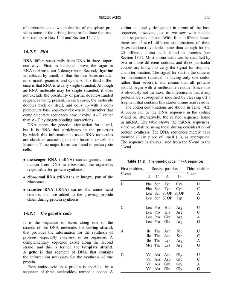
The codon combinations are shown in Table 14.2.
A codon can be the DNA sequence in the coding
strand or, alternatively, the related sequence found
in mRNA. The table shows the mRNA sequences,
since we shall be using these during consideration of
protein synthesis. The DNA sequences merely have
thymine (T) in place of uracil (U), as appropriate.
The sequence is always listed from the 5
-end to the
3
-end.
Table 14.2 The genetic code: mRNA sequences
First position,
Second position
Third position,
5
-end
U C
A
G
3
-end
U
Phe Ser Tyr
Cys
U
Phe Ser Tyr
Cys
C
Leu Ser STOP STOP
A
Leu Ser STOP Trp
G
C
L e uP r o H i s
A r g
U
Leu Pro His
Arg
C
Leu Pro Gln
Arg
A
Leu Pro Gln
Arg
G
A
I l e T h r A s n
S e r
U
Ile Thr Asn
Ser
C
Ile Thr Lys
Arg
A
Met Thr Lys
Arg
G
G
Val Ala Asp Gly
U
Val Ala Asp Gly
C
Val Ala Glu
Gly
A
Val Ala Glu
Gly
G
555
of diphosphate to two molecules of phosphate provides some of the driving force to facilitate the reaction (compare Box 14.3 and Section 15.4.1).
14.2.3 RNA
RNA differs structurally from DNA in three important ways. First, as indicated above, the sugar in
RNA is ribose, not 2-deoxyribose. Second, thymine
is replaced by uracil, so that the four bases are adenine, uracil, guanine, and cytosine. The third difference is that RNA is usually single stranded. Although
an RNA molecule may be single stranded, it does
not exclude the possibility of partial double-stranded
sequences being present. In such cases, the molecule
doubles back on itself, and coils up with a complementary base sequence elsewhere. Remember that
complementary sequences now involve A–U rather
than A–T hydrogen-bonding interactions.
DNA stores the genetic information for a cell,
but it is RNA that participates in the processes
by which this information is used. RNA molecules
are classified according to their function or cellular
location. Three major forms are found in prokaryotic
cells:
• messenger RNA (mRNA) carries genetic information from DNA to ribosomes, the organelles
responsible for protein synthesis;
• ribosomal RNA (rRNA) is an integral part of the
ribosomes;
• transfer RNA (tRNA) carries the amino acid
residues that are added to the growing peptide
chain during protein synthesis.
14.2.4 The genetic code
It is the sequence of bases along one of the
strands of the DNA molecule, the coding strand,
that provides the information for the synthesis of
proteins, especially enzymes, in an organism. A
complementary sequence exists along the second
strand, and this is termed the template strand.
A gene is that segment of DNA that contains
the information necessary for the synthesis of one
protein.
Each amino acid in a protein is specified by a
sequence of three nucleotides, termed a codon. A
codon is usually designated in terms of the base
sequence, however, just as we saw with nucleic
acid sequences above. With four different bases,
there are 4
3
= 64 different combinations of three
bases (codons) available, more than enough for the
20 different amino acids found in proteins (see
Section 13.1). Most amino acids can be specified by
two or more different codons, and three particular
codons are known to carry the signal for stop, i.e.
chain termination. The signal for start is the same as
for methionine (unusual in having only one codon
rather than several), and means that all proteins
should begin with a methionine residue. Since this
is obviously not the case, the inference is that many
proteins are subsequently modified by cleaving off a
fragment that contains this starter amino acid residue.
The codon combinations are shown in Table 14.2.
A codon can be the DNA sequence in the coding
strand or, alternatively, the related sequence found
in mRNA. The table shows the mRNA sequences,
since we shall be using these during consideration of
protein synthesis. The DNA sequences merely have
thymine (T) in place of uracil (U), as appropriate.
The sequence is always listed from the 5
-end to the
3
-end.
Table 14.2 The genetic code: mRNA sequences
First position,
Second position
Third position,
5
-end
U C
A
G
3
-end
U
Phe Ser Tyr
Cys
U
Phe Ser Tyr
Cys
C
Leu Ser STOP STOP
A
Leu Ser STOP Trp
G
C
L e uP r o H i s
A r g
U
Leu Pro His
Arg
C
Leu Pro Gln
Arg
A
Leu Pro Gln
Arg
G
A
I l e T h r A s n
S e r
U
Ile Thr Asn
Ser
C
Ile Thr Lys
Arg
A
Met Thr Lys
Arg
G
G
Val Ala Asp Gly
U
Val Ala Asp Gly
C
Val Ala Glu
Gly
A
Val Ala Glu
Gly
G