

46
M. Stein
4.2 Model Complexes
Structural and functional model complexes are valuable systems to gain insight into
the structure and electronic and spectroscopic properties of these small systems. Very
often, model complexes are available in larger amount and higher purity compared
to enzymatic systems. Thus, structural data (from e.g. X-ray crystallography) and
magnetic resonance data are more facile to obtain and may be analyzed in more
detail. The spectroscopic and computational results obtained for the biomimetic or
bioinspired model may serve as a benchmark or comparison with an enzymatic
system. In model compounds, magnetic and structural symmetry axes are very often
coincident since both ligand field and magnetic field splittings are determined by
ligand strength and coordination geometry.
4.2.1 Transition Metal Single Crystal EPR
In bis(malenonitriledithiolato)nickelate(III) (Ni(mnt)
−
2 ) the nickel atom possesses a
first coordination sphere similar to that of the [NiFe]-hydrogenase enzyme. Nickel
is in the +3 oxidation state as in the Ni–A, Ni–B, and Ni–C states in the enzyme.
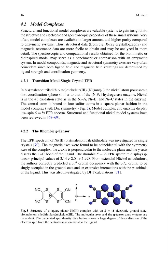
The central atom is bound to four sulfur atoms in a square-planar fashion in the
model complex (with D 2h symmetry) (Fig. 5). Model complex and enzyme display
low-spin S ½ EPR spectra. Structural and functional nickel model systems have
been reviewed in [67–69].
4.2.2 The Rhombic g-Tensor
The EPR spectrum of Ni(III) bis(malenonitrile)dithiolate was investigated in single
crystals [70]. The magnetic axes were found to be coincidental with the symmetry
axes of the complex: the z-axis is perpendicular to the molecule plane and the y-axis
bisects the C=C bond of the ligand. The rhombic S ½ EPR spectrum displays gtensor principal values of 2.14 > 2.04 > 1.998. From extended Hückel calculations,
the authors correctly predicted a 3d
7 orbital occupancy with the 3d yz orbital to be
singly occupied in the ground state and an extensive interactions with the π-orbitals
of the ligand. This was also investigated by DFT calculations [71].
Fig. 5 Structure of a square-planar Ni(III) complex with an S ½ electronic ground state:
bis(malenonitriledithiolato)nickelate(III). The molecular axes and the g-tensor axes systems are
coincident. The calculated spin density distribution shows a large degree of delocalization of the
electron spin from the central transition metal to the ligand
M. Stein
4.2 Model Complexes
Structural and functional model complexes are valuable systems to gain insight into
the structure and electronic and spectroscopic properties of these small systems. Very
often, model complexes are available in larger amount and higher purity compared
to enzymatic systems. Thus, structural data (from e.g. X-ray crystallography) and
magnetic resonance data are more facile to obtain and may be analyzed in more
detail. The spectroscopic and computational results obtained for the biomimetic or
bioinspired model may serve as a benchmark or comparison with an enzymatic
system. In model compounds, magnetic and structural symmetry axes are very often
coincident since both ligand field and magnetic field splittings are determined by
ligand strength and coordination geometry.
4.2.1 Transition Metal Single Crystal EPR
In bis(malenonitriledithiolato)nickelate(III) (Ni(mnt)
−
2 ) the nickel atom possesses a
first coordination sphere similar to that of the [NiFe]-hydrogenase enzyme. Nickel
is in the +3 oxidation state as in the Ni–A, Ni–B, and Ni–C states in the enzyme.
The central atom is bound to four sulfur atoms in a square-planar fashion in the
model complex (with D 2h symmetry) (Fig. 5). Model complex and enzyme display
low-spin S ½ EPR spectra. Structural and functional nickel model systems have
been reviewed in [67–69].
4.2.2 The Rhombic g-Tensor
The EPR spectrum of Ni(III) bis(malenonitrile)dithiolate was investigated in single
crystals [70]. The magnetic axes were found to be coincidental with the symmetry
axes of the complex: the z-axis is perpendicular to the molecule plane and the y-axis
bisects the C=C bond of the ligand. The rhombic S ½ EPR spectrum displays gtensor principal values of 2.14 > 2.04 > 1.998. From extended Hückel calculations,
the authors correctly predicted a 3d
7 orbital occupancy with the 3d yz orbital to be
singly occupied in the ground state and an extensive interactions with the π-orbitals
of the ligand. This was also investigated by DFT calculations [71].
Fig. 5 Structure of a square-planar Ni(III) complex with an S ½ electronic ground state:
bis(malenonitriledithiolato)nickelate(III). The molecular axes and the g-tensor axes systems are
coincident. The calculated spin density distribution shows a large degree of delocalization of the
electron spin from the central transition metal to the ligand