

How Metal Coordination in the Ca-, Ce- and Eu-Containing …
495
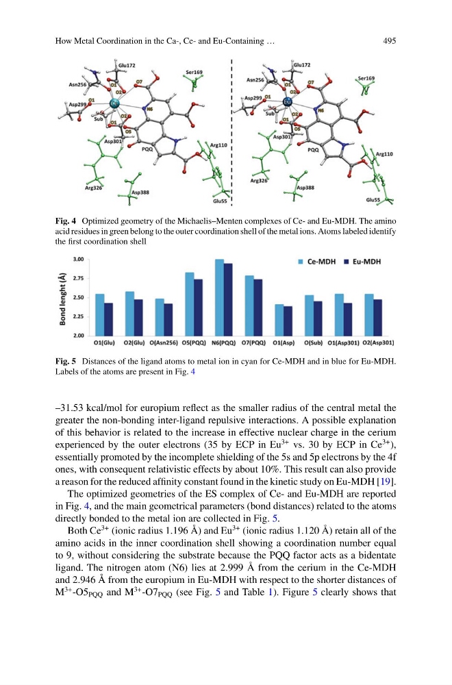
Fig. 4 Optimized geometry of the Michaelis–Menten complexes of Ce- and Eu-MDH. The amino
acid residues in green belong to the outer coordination shell of the metal ions. Atoms labeled identify
the first coordination shell
Fig. 5 Distances of the ligand atoms to metal ion in cyan for Ce-MDH and in blue for Eu-MDH.
Labels of the atoms are present in Fig. 4
–31.53 kcal/mol for europium reflect as the smaller radius of the central metal the
greater the non-bonding inter-ligand repulsive interactions. A possible explanation
of this behavior is related to the increase in effective nuclear charge in the cerium
experienced by the outer electrons (35 by ECP in Eu
3+ vs. 30 by ECP in Ce
3+ ),
essentially promoted by the incomplete shielding of the 5s and 5p electrons by the 4f
ones, with consequent relativistic effects by about 10%. This result can also provide
a reason for the reduced affinity constant found in the kinetic study on Eu-MDH [19].
The optimized geometries of the ES complex of Ce- and Eu-MDH are reported
in Fig. 4, and the main geometrical parameters (bond distances) related to the atoms
directly bonded to the metal ion are collected in Fig. 5.
Both Ce
3+ (ionic radius 1.196 Å) and Eu
3+ (ionic radius 1.120 Å) retain all of the
amino acids in the inner coordination shell showing a coordination number equal
to 9, without considering the substrate because the PQQ factor acts as a bidentate
ligand. The nitrogen atom (N6) lies at 2.999 Å from the cerium in the Ce-MDH
and 2.946 Å from the europium in Eu-MDH with respect to the shorter distances of
M
3+ -O5 PQQ and M
3+ -O7 PQQ (see Fig. 5 and Table 1). Figure 5 clearly shows that
495
Fig. 4 Optimized geometry of the Michaelis–Menten complexes of Ce- and Eu-MDH. The amino
acid residues in green belong to the outer coordination shell of the metal ions. Atoms labeled identify
the first coordination shell
Fig. 5 Distances of the ligand atoms to metal ion in cyan for Ce-MDH and in blue for Eu-MDH.
Labels of the atoms are present in Fig. 4
–31.53 kcal/mol for europium reflect as the smaller radius of the central metal the
greater the non-bonding inter-ligand repulsive interactions. A possible explanation
of this behavior is related to the increase in effective nuclear charge in the cerium
experienced by the outer electrons (35 by ECP in Eu
3+ vs. 30 by ECP in Ce
3+ ),
essentially promoted by the incomplete shielding of the 5s and 5p electrons by the 4f
ones, with consequent relativistic effects by about 10%. This result can also provide
a reason for the reduced affinity constant found in the kinetic study on Eu-MDH [19].
The optimized geometries of the ES complex of Ce- and Eu-MDH are reported
in Fig. 4, and the main geometrical parameters (bond distances) related to the atoms
directly bonded to the metal ion are collected in Fig. 5.
Both Ce
3+ (ionic radius 1.196 Å) and Eu
3+ (ionic radius 1.120 Å) retain all of the
amino acids in the inner coordination shell showing a coordination number equal
to 9, without considering the substrate because the PQQ factor acts as a bidentate
ligand. The nitrogen atom (N6) lies at 2.999 Å from the cerium in the Ce-MDH
and 2.946 Å from the europium in Eu-MDH with respect to the shorter distances of
M
3+ -O5 PQQ and M
3+ -O7 PQQ (see Fig. 5 and Table 1). Figure 5 clearly shows that