

Computational Versus Experimental Spectroscopy …
167
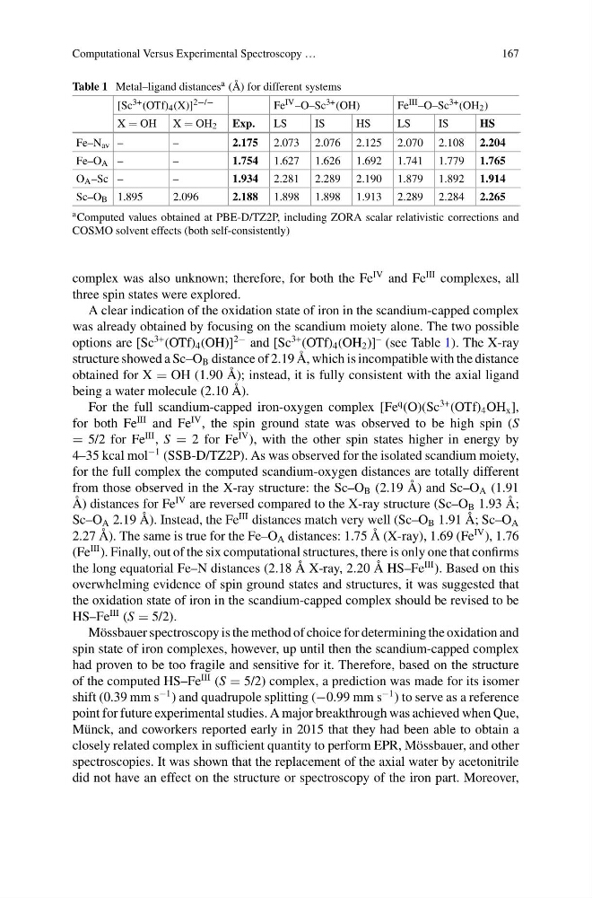
Table 1 Metal–ligand distances a (Å) for different systems
[Sc 3+ (OTf) 4 (X)] 2−/−
Fe IV –O–Sc 3+ (OH)
Fe III –O–Sc 3+ (OH 2 )
X OH X OH 2 Exp.
LS
IS
HS
LS
IS
HS
Fe–N av –
–
2.175 2.073 2.076 2.125 2.070 2.108 2.204
Fe–O A –
–
1.754 1.627 1.626 1.692 1.741 1.779 1.765
O A –Sc –
–
1.934 2.281 2.289 2.190 1.879 1.892 1.914
Sc–O B 1.895
2.096
2.188 1.898 1.898 1.913 2.289 2.284 2.265
a Computed values obtained at PBE-D/TZ2P, including ZORA scalar relativistic corrections and
COSMO solvent effects (both self-consistently)
complex was also unknown; therefore, for both the Fe
IV and Fe
III complexes, all
three spin states were explored.
A clear indication of the oxidation state of iron in the scandium-capped complex
was already obtained by focusing on the scandium moiety alone. The two possible
options are [Sc
3+ (OTf) 4 (OH)]
2− and [Sc
3+ (OTf) 4 (OH 2 )]
– (see Table 1). The X-ray
structure showed a Sc–O B distance of 2.19 Å, which is incompatible with the distance
obtained for X OH (1.90 Å); instead, it is fully consistent with the axial ligand
being a water molecule (2.10 Å).
For the full scandium-capped iron-oxygen complex [Fe
q (O)(Sc
3+ (OTf) 4 OH x ],
for both Fe
III and Fe
IV , the spin ground state was observed to be high spin (S
5/2 for Fe
III , S 2 for Fe
IV ), with the other spin states higher in energy by
4–35 kcal mol
−1 (SSB-D/TZ2P). As was observed for the isolated scandium moiety,
for the full complex the computed scandium-oxygen distances are totally different
from those observed in the X-ray structure: the Sc–O B (2.19 Å) and Sc–O A (1.91
Å) distances for Fe
IV are reversed compared to the X-ray structure (Sc–O B 1.93 Å;
Sc–O A 2.19 Å). Instead, the Fe
III distances match very well (Sc–O B 1.91 Å; Sc–O A
2.27 Å). The same is true for the Fe–O A distances: 1.75 Å (X-ray), 1.69 (Fe
IV ), 1.76
(Fe
III ). Finally, out of the six computational structures, there is only one that confirms
the long equatorial Fe–N distances (2.18 Å X-ray, 2.20 Å HS–Fe
III ). Based on this
overwhelming evidence of spin ground states and structures, it was suggested that
the oxidation state of iron in the scandium-capped complex should be revised to be
HS–Fe
III (S 5/2).
Mössbauer spectroscopy is the method of choice for determining the oxidation and
spin state of iron complexes, however, up until then the scandium-capped complex
had proven to be too fragile and sensitive for it. Therefore, based on the structure
of the computed HS–Fe
III (S 5/2) complex, a prediction was made for its isomer
shift (0.39 mm s
−1 ) and quadrupole splitting (−0.99 mm s
−1 ) to serve as a reference
point for future experimental studies. A major breakthrough was achieved when Que,
Münck, and coworkers reported early in 2015 that they had been able to obtain a
closely related complex in sufficient quantity to perform EPR, Mössbauer, and other
spectroscopies. It was shown that the replacement of the axial water by acetonitrile
did not have an effect on the structure or spectroscopy of the iron part. Moreover,
167
Table 1 Metal–ligand distances a (Å) for different systems
[Sc 3+ (OTf) 4 (X)] 2−/−
Fe IV –O–Sc 3+ (OH)
Fe III –O–Sc 3+ (OH 2 )
X OH X OH 2 Exp.
LS
IS
HS
LS
IS
HS
Fe–N av –
–
2.175 2.073 2.076 2.125 2.070 2.108 2.204
Fe–O A –
–
1.754 1.627 1.626 1.692 1.741 1.779 1.765
O A –Sc –
–
1.934 2.281 2.289 2.190 1.879 1.892 1.914
Sc–O B 1.895
2.096
2.188 1.898 1.898 1.913 2.289 2.284 2.265
a Computed values obtained at PBE-D/TZ2P, including ZORA scalar relativistic corrections and
COSMO solvent effects (both self-consistently)
complex was also unknown; therefore, for both the Fe
IV and Fe
III complexes, all
three spin states were explored.
A clear indication of the oxidation state of iron in the scandium-capped complex
was already obtained by focusing on the scandium moiety alone. The two possible
options are [Sc
3+ (OTf) 4 (OH)]
2− and [Sc
3+ (OTf) 4 (OH 2 )]
– (see Table 1). The X-ray
structure showed a Sc–O B distance of 2.19 Å, which is incompatible with the distance
obtained for X OH (1.90 Å); instead, it is fully consistent with the axial ligand
being a water molecule (2.10 Å).
For the full scandium-capped iron-oxygen complex [Fe
q (O)(Sc
3+ (OTf) 4 OH x ],
for both Fe
III and Fe
IV , the spin ground state was observed to be high spin (S
5/2 for Fe
III , S 2 for Fe
IV ), with the other spin states higher in energy by
4–35 kcal mol
−1 (SSB-D/TZ2P). As was observed for the isolated scandium moiety,
for the full complex the computed scandium-oxygen distances are totally different
from those observed in the X-ray structure: the Sc–O B (2.19 Å) and Sc–O A (1.91
Å) distances for Fe
IV are reversed compared to the X-ray structure (Sc–O B 1.93 Å;
Sc–O A 2.19 Å). Instead, the Fe
III distances match very well (Sc–O B 1.91 Å; Sc–O A
2.27 Å). The same is true for the Fe–O A distances: 1.75 Å (X-ray), 1.69 (Fe
IV ), 1.76
(Fe
III ). Finally, out of the six computational structures, there is only one that confirms
the long equatorial Fe–N distances (2.18 Å X-ray, 2.20 Å HS–Fe
III ). Based on this
overwhelming evidence of spin ground states and structures, it was suggested that
the oxidation state of iron in the scandium-capped complex should be revised to be
HS–Fe
III (S 5/2).
Mössbauer spectroscopy is the method of choice for determining the oxidation and
spin state of iron complexes, however, up until then the scandium-capped complex
had proven to be too fragile and sensitive for it. Therefore, based on the structure
of the computed HS–Fe
III (S 5/2) complex, a prediction was made for its isomer
shift (0.39 mm s
−1 ) and quadrupole splitting (−0.99 mm s
−1 ) to serve as a reference
point for future experimental studies. A major breakthrough was achieved when Que,
Münck, and coworkers reported early in 2015 that they had been able to obtain a
closely related complex in sufficient quantity to perform EPR, Mössbauer, and other
spectroscopies. It was shown that the replacement of the axial water by acetonitrile
did not have an effect on the structure or spectroscopy of the iron part. Moreover,