

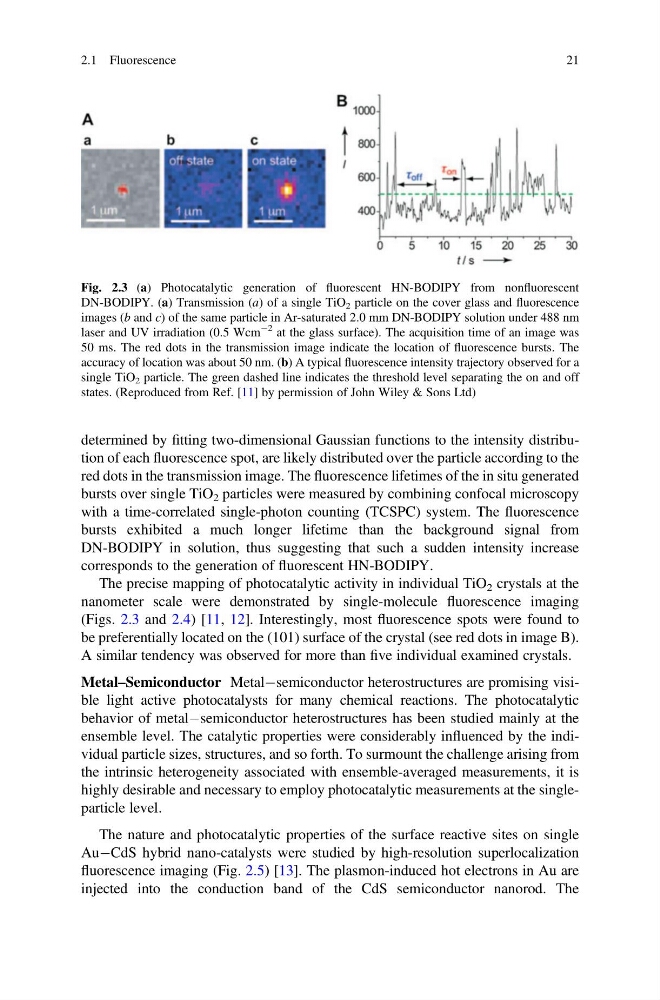
determined by fitting two-dimensional Gaussian functions to the intensity distribution of each fluorescence spot, are likely distributed over the particle according to the
red dots in the transmission image. The fluorescence lifetimes of the in situ generated
bursts over single TiO 2 particles were measured by combining confocal microscopy
with a time-correlated single-photon counting (TCSPC) system. The fluorescence
bursts exhibited a much longer lifetime than the background signal from
DN-BODIPY in solution, thus suggesting that such a sudden intensity increase
corresponds to the generation of fluorescent HN-BODIPY.
The precise mapping of photocatalytic activity in individual TiO 2 crystals at the
nanometer scale were demonstrated by single-molecule fluorescence imaging
(Figs. 2.3 and 2.4) [11, 12]. Interestingly, most fluorescence spots were found to
be preferentially located on the (101) surface of the crystal (see red dots in image B).
A similar tendency was observed for more than five individual examined crystals.
Metal–Semiconductor MetalÀsemiconductor heterostructures are promising visible light active photocatalysts for many chemical reactions. The photocatalytic
behavior of metalÀsemiconductor heterostructures has been studied mainly at the
ensemble level. The catalytic properties were considerably influenced by the individual particle sizes, structures, and so forth. To surmount the challenge arising from
the intrinsic heterogeneity associated with ensemble-averaged measurements, it is
highly desirable and necessary to employ photocatalytic measurements at the singleparticle level.
The nature and photocatalytic properties of the surface reactive sites on single
AuÀCdS hybrid nano-catalysts were studied by high-resolution superlocalization
fluorescence imaging (Fig. 2.5) [13]. The plasmon-induced hot electrons in Au are
injected into the conduction band of the CdS semiconductor nanorod. The
Fig. 2.3 (a) Photocatalytic generation of fluorescent HN-BODIPY from nonfluorescent
DN-BODIPY. (a) Transmission (a) of a single TiO 2 particle on the cover glass and fluorescence
images (b and c) of the same particle in Ar-saturated 2.0 mm DN-BODIPY solution under 488 nm
laser and UV irradiation (0.5 Wcm
À2 at the glass surface). The acquisition time of an image was
50 ms. The red dots in the transmission image indicate the location of fluorescence bursts. The
accuracy of location was about 50 nm. (b) A typical fluorescence intensity trajectory observed for a
single TiO 2 particle. The green dashed line indicates the threshold level separating the on and off
states. (Reproduced from Ref. [11] by permission of John Wiley & Sons Ltd)
2.1 Fluorescence
21
red dots in the transmission image. The fluorescence lifetimes of the in situ generated
bursts over single TiO 2 particles were measured by combining confocal microscopy
with a time-correlated single-photon counting (TCSPC) system. The fluorescence
bursts exhibited a much longer lifetime than the background signal from
DN-BODIPY in solution, thus suggesting that such a sudden intensity increase
corresponds to the generation of fluorescent HN-BODIPY.
The precise mapping of photocatalytic activity in individual TiO 2 crystals at the
nanometer scale were demonstrated by single-molecule fluorescence imaging
(Figs. 2.3 and 2.4) [11, 12]. Interestingly, most fluorescence spots were found to
be preferentially located on the (101) surface of the crystal (see red dots in image B).
A similar tendency was observed for more than five individual examined crystals.
Metal–Semiconductor MetalÀsemiconductor heterostructures are promising visible light active photocatalysts for many chemical reactions. The photocatalytic
behavior of metalÀsemiconductor heterostructures has been studied mainly at the
ensemble level. The catalytic properties were considerably influenced by the individual particle sizes, structures, and so forth. To surmount the challenge arising from
the intrinsic heterogeneity associated with ensemble-averaged measurements, it is
highly desirable and necessary to employ photocatalytic measurements at the singleparticle level.
The nature and photocatalytic properties of the surface reactive sites on single
AuÀCdS hybrid nano-catalysts were studied by high-resolution superlocalization
fluorescence imaging (Fig. 2.5) [13]. The plasmon-induced hot electrons in Au are
injected into the conduction band of the CdS semiconductor nanorod. The
Fig. 2.3 (a) Photocatalytic generation of fluorescent HN-BODIPY from nonfluorescent
DN-BODIPY. (a) Transmission (a) of a single TiO 2 particle on the cover glass and fluorescence
images (b and c) of the same particle in Ar-saturated 2.0 mm DN-BODIPY solution under 488 nm
laser and UV irradiation (0.5 Wcm
À2 at the glass surface). The acquisition time of an image was
50 ms. The red dots in the transmission image indicate the location of fluorescence bursts. The
accuracy of location was about 50 nm. (b) A typical fluorescence intensity trajectory observed for a
single TiO 2 particle. The green dashed line indicates the threshold level separating the on and off
states. (Reproduced from Ref. [11] by permission of John Wiley & Sons Ltd)
2.1 Fluorescence
21