

techniques, Datye et al. [129] put forth distinguishing views against Bickley et al.
[76]. The characterization results showed that the mixed-phase P25 was composed
of individual single-crystal particles of anatase and rutile rather than anatase particles
covered by a layer of rutile. Subsequently, their view was confirmed by Zhang et al.
[130] and Ohno et al. [131]. But they did not further study the mechanism of the
enhanced photocatalytic performance of the mixed-phase TiO 2 .
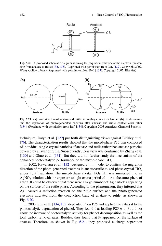
In 2002, Kawahara et al. [132] designed a film model to confirm the migration
direction of the photo-generated excitons in anatase/rutile mixed-phase crystal TiO 2
under light irradiation. The mixed-phase crystal TiO 2 film was immersed into an
AgNO 3 solution with the exposure to light over a period of time at the atmosphere of
argon. It could be observed that there were a large number of Ag particles appearing
on the surface of the rutile phase. According to the phenomenon, they inferred that
Ag
+ caused a reduction reaction on the rutile surface and the photo-generated
electrons migrated from the conduction band of anatase to rutile, as shown in
Fig. 6.20.
In 2003, Sun et al. [134, 135] deposited Pt on P25 and applied the catalyst to the
photocatalytic degradation of phenol. They found that loading P25 with Pt did not
show the increase of photocatalytic activity for phenol decomposition as well as the
total carbon removal rates. Besides, they found that Pt appeared on the surface of
anatase. Therefore, as shown in Fig. 6.21, they proposed a charge separation
Fig. 6.20 A proposed schematic diagram showing the migration behavior of the electron transferring from anatase to rutile [132, 133]. (Reprinted with permission from Ref. [132]. Copyright 2002,
Wiley Online Library. Reprinted with permission from Ref. [133]. Copyright 2007, Elsevier)
E FA
E FR
VB
hv
hv
VB
CB
CB
O 2
O 2
H 2 O
OH
–
OH
•
3.2 eV
3.0 eV
Anatase
Anatase
Rutile
Rutile
(a)
(b)
Fig. 6.21 (a) Band structure of anatase and rutile before they contact each other; (b) band structure
and the separation of photo-generated excitons after anatase and rutile contact each other
[134]. (Reprinted with permission from Ref. [134]. Copyright 2003 American Chemical Society)
162
6 Phase Control of TiO 2 Photocatalyst
[76]. The characterization results showed that the mixed-phase P25 was composed
of individual single-crystal particles of anatase and rutile rather than anatase particles
covered by a layer of rutile. Subsequently, their view was confirmed by Zhang et al.
[130] and Ohno et al. [131]. But they did not further study the mechanism of the
enhanced photocatalytic performance of the mixed-phase TiO 2 .
In 2002, Kawahara et al. [132] designed a film model to confirm the migration
direction of the photo-generated excitons in anatase/rutile mixed-phase crystal TiO 2
under light irradiation. The mixed-phase crystal TiO 2 film was immersed into an
AgNO 3 solution with the exposure to light over a period of time at the atmosphere of
argon. It could be observed that there were a large number of Ag particles appearing
on the surface of the rutile phase. According to the phenomenon, they inferred that
Ag
+ caused a reduction reaction on the rutile surface and the photo-generated
electrons migrated from the conduction band of anatase to rutile, as shown in
Fig. 6.20.
In 2003, Sun et al. [134, 135] deposited Pt on P25 and applied the catalyst to the
photocatalytic degradation of phenol. They found that loading P25 with Pt did not
show the increase of photocatalytic activity for phenol decomposition as well as the
total carbon removal rates. Besides, they found that Pt appeared on the surface of
anatase. Therefore, as shown in Fig. 6.21, they proposed a charge separation
Fig. 6.20 A proposed schematic diagram showing the migration behavior of the electron transferring from anatase to rutile [132, 133]. (Reprinted with permission from Ref. [132]. Copyright 2002,
Wiley Online Library. Reprinted with permission from Ref. [133]. Copyright 2007, Elsevier)
E FA
E FR
VB
hv
hv
VB
CB
CB
O 2
O 2
H 2 O
OH
–
OH
•
3.2 eV
3.0 eV
Anatase
Anatase
Rutile
Rutile
(a)
(b)
Fig. 6.21 (a) Band structure of anatase and rutile before they contact each other; (b) band structure
and the separation of photo-generated excitons after anatase and rutile contact each other
[134]. (Reprinted with permission from Ref. [134]. Copyright 2003 American Chemical Society)
162
6 Phase Control of TiO 2 Photocatalyst