

mesoporous TiO 2 nanospindles. Then following the same procedure, the TiO 2
nanospindles were chemically anchored onto the graphene oxide (GO) to form a
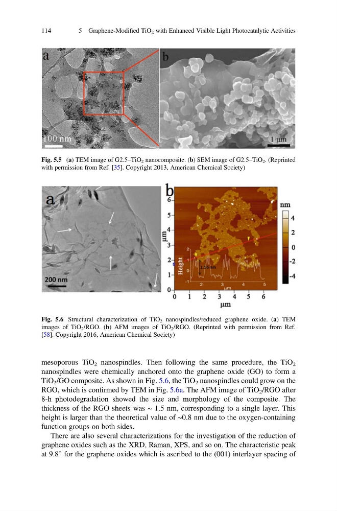
TiO 2 /GO composite. As shown in Fig. 5.6, the TiO 2 nanospindles could grow on the
RGO, which is confirmed by TEM in Fig. 5.6a. The AFM image of TiO 2 /RGO after
8-h photodegradation showed the size and morphology of the composite. The
thickness of the RGO sheets was ~ 1.5 nm, corresponding to a single layer. This
height is larger than the theoretical value of ~0.8 nm due to the oxygen-containing
function groups on both sides.
There are also several characterizations for the investigation of the reduction of
graphene oxides such as the XRD, Raman, XPS, and so on. The characteristic peak
at 9.8
for the graphene oxides which is ascribed to the (001) interlayer spacing of
Fig. 5.5 (a) TEM image of G2.5–TiO 2 nanocomposite. (b) SEM image of G2.5–TiO 2 . (Reprinted
with permission from Ref. [35]. Copyright 2013, American Chemical Society)
Fig. 5.6 Structural characterization of TiO 2 nanospindles/reduced graphene oxide. (a) TEM
images of TiO 2 /RGO. (b) AFM images of TiO 2 /RGO. (Reprinted with permission from Ref.
[58]. Copyright 2016, American Chemical Society)
114
5 Graphene-Modified TiO 2 with Enhanced Visible Light Photocatalytic Activities
nanospindles were chemically anchored onto the graphene oxide (GO) to form a
TiO 2 /GO composite. As shown in Fig. 5.6, the TiO 2 nanospindles could grow on the
RGO, which is confirmed by TEM in Fig. 5.6a. The AFM image of TiO 2 /RGO after
8-h photodegradation showed the size and morphology of the composite. The
thickness of the RGO sheets was ~ 1.5 nm, corresponding to a single layer. This
height is larger than the theoretical value of ~0.8 nm due to the oxygen-containing
function groups on both sides.
There are also several characterizations for the investigation of the reduction of
graphene oxides such as the XRD, Raman, XPS, and so on. The characteristic peak
at 9.8
for the graphene oxides which is ascribed to the (001) interlayer spacing of
Fig. 5.5 (a) TEM image of G2.5–TiO 2 nanocomposite. (b) SEM image of G2.5–TiO 2 . (Reprinted
with permission from Ref. [35]. Copyright 2013, American Chemical Society)
Fig. 5.6 Structural characterization of TiO 2 nanospindles/reduced graphene oxide. (a) TEM
images of TiO 2 /RGO. (b) AFM images of TiO 2 /RGO. (Reprinted with permission from Ref.
[58]. Copyright 2016, American Chemical Society)
114
5 Graphene-Modified TiO 2 with Enhanced Visible Light Photocatalytic Activities