

(X standing for F, Cl and Br). It is clearly seen that F is more strongly bonded than
Cl and Br. This result is in agreement with their binding energies, which are
−632.8, −571.0 and −562.6 kcal/mol, respectively.
18.7.5.2 Zn
2+ Fingers
Metals play a decisive role in many protein active sites as cofactors. The zinc finger
is a small protein structural motif that can be found in many biological systems. It is
characterized by the fact than one or more zinc ions can stabilize the fold of a
protein. One or two Zn(II) cations [100] are often tetrahedrally coordinated to four
or six amino acids such as cysteine (Cys) or histidine (His) forming four major
cores: ZnCys 4 , ZnCys 3 His, ZnCys 2 His 2 and ZnCys 6 [101].
Starting from the ZnCys 4 core, its modeling can be carried out by substitution of
Cys by methyl thiolate, CH 3 S
– [101]. In order to analyze the effect of the environement within the HSAB theory, we have analyzed the series M
2 þ
½SCH 3
2À
4 ,
with M = Mg, Zn, Pd (from hard to soft Lewis acids). All complexes were optimized at the ωB3LYP/6-31++G** level of theory except for Pd, which was optimized with the pseudopotential LANL2DZ.
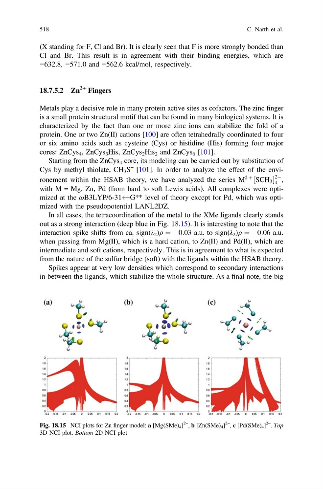
In all cases, the tetracoordination of the metal to the XMe ligands clearly stands
out as a strong interaction (deep blue in Fig. 18.15). It is interesting to note that the
interaction spike shifts from ca. sign(λ 2 )q ¼ À0:03 a.u. to sign(λ 2 )q ¼ À0:06 a.u.
when passing from Mg(II), which is a hard cation, to Zn(II) and Pd(II), which are
intermediate and soft cations, respectively. This is in agreement to what is expected
from the nature of the sulfur bridge (soft) with the ligands within the HSAB theory.
Spikes appear at very low densities which correspond to secondary interactions
in between the ligands, which stabilize the whole structure. As a final note, the big
Fig. 18.15 NCI plots for Zn finger model: a [Mg(SMe) 4 ]
2–
, b [Zn(SMe) 4 ]
2–
, c [Pd(SMe) 4 ]
2–
. Top
3D NCI plot. Bottom 2D NCI plot
518
C. Narth et al.
Cl and Br. This result is in agreement with their binding energies, which are
−632.8, −571.0 and −562.6 kcal/mol, respectively.
18.7.5.2 Zn
2+ Fingers
Metals play a decisive role in many protein active sites as cofactors. The zinc finger
is a small protein structural motif that can be found in many biological systems. It is
characterized by the fact than one or more zinc ions can stabilize the fold of a
protein. One or two Zn(II) cations [100] are often tetrahedrally coordinated to four
or six amino acids such as cysteine (Cys) or histidine (His) forming four major
cores: ZnCys 4 , ZnCys 3 His, ZnCys 2 His 2 and ZnCys 6 [101].
Starting from the ZnCys 4 core, its modeling can be carried out by substitution of
Cys by methyl thiolate, CH 3 S
– [101]. In order to analyze the effect of the environement within the HSAB theory, we have analyzed the series M
2 þ
½SCH 3
2À
4 ,
with M = Mg, Zn, Pd (from hard to soft Lewis acids). All complexes were optimized at the ωB3LYP/6-31++G** level of theory except for Pd, which was optimized with the pseudopotential LANL2DZ.
In all cases, the tetracoordination of the metal to the XMe ligands clearly stands
out as a strong interaction (deep blue in Fig. 18.15). It is interesting to note that the
interaction spike shifts from ca. sign(λ 2 )q ¼ À0:03 a.u. to sign(λ 2 )q ¼ À0:06 a.u.
when passing from Mg(II), which is a hard cation, to Zn(II) and Pd(II), which are
intermediate and soft cations, respectively. This is in agreement to what is expected
from the nature of the sulfur bridge (soft) with the ligands within the HSAB theory.
Spikes appear at very low densities which correspond to secondary interactions
in between the ligands, which stabilize the whole structure. As a final note, the big
Fig. 18.15 NCI plots for Zn finger model: a [Mg(SMe) 4 ]
2–
, b [Zn(SMe) 4 ]
2–
, c [Pd(SMe) 4 ]
2–
. Top
3D NCI plot. Bottom 2D NCI plot
518
C. Narth et al.