

14
A. Monari and X. Assfeld
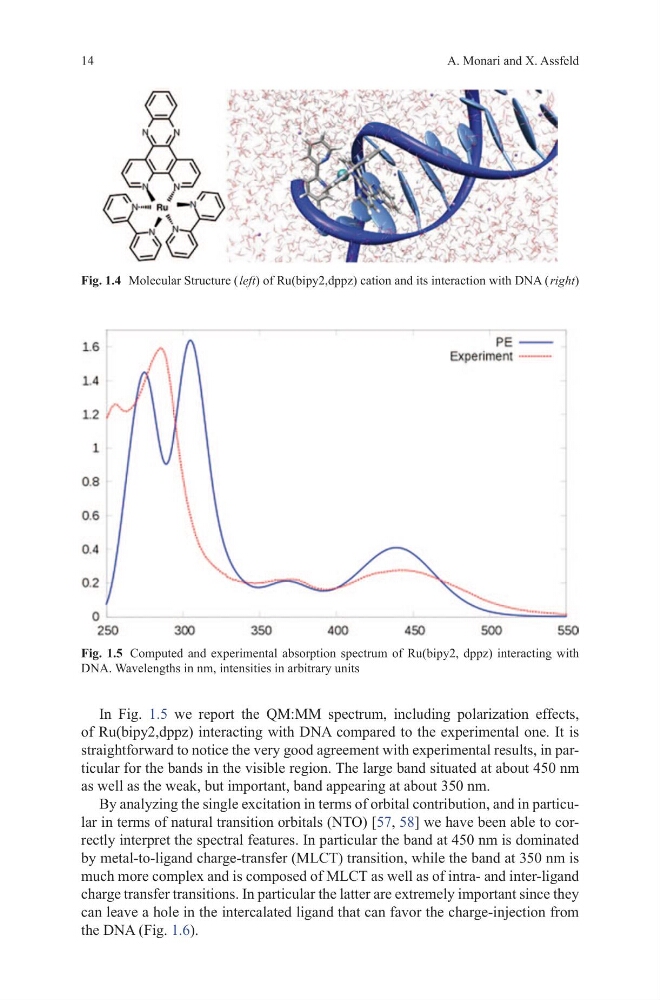
In Fig. 1.5 we report the QM:MM spectrum, including polarization effects,
of Ru(bipy2,dppz) interacting with DNA compared to the experimental one. It is
straightforward to notice the very good agreement with experimental results, in particular for the bands in the visible region. The large band situated at about 450 nm
as well as the weak, but important, band appearing at about 350 nm.
By analyzing the single excitation in terms of orbital contribution, and in particular in terms of natural transition orbitals (NTO) [57, 58] we have been able to correctly interpret the spectral features. In particular the band at 450 nm is dominated
by metal-to-ligand charge-transfer (MLCT) transition, while the band at 350 nm is
much more complex and is composed of MLCT as well as of intra- and inter-ligand
charge transfer transitions. In particular the latter are extremely important since they
can leave a hole in the intercalated ligand that can favor the charge-injection from
the DNA (Fig. 1.6).
Fig. 1.4 Molecular Structure ( left) of Ru(bipy2,dppz) cation and its interaction with DNA ( right)
Fig. 1.5 Computed and experimental absorption spectrum of Ru(bipy2, dppz) interacting with
DNA. Wavelengths in nm, intensities in arbitrary units
A. Monari and X. Assfeld
In Fig. 1.5 we report the QM:MM spectrum, including polarization effects,
of Ru(bipy2,dppz) interacting with DNA compared to the experimental one. It is
straightforward to notice the very good agreement with experimental results, in particular for the bands in the visible region. The large band situated at about 450 nm
as well as the weak, but important, band appearing at about 350 nm.
By analyzing the single excitation in terms of orbital contribution, and in particular in terms of natural transition orbitals (NTO) [57, 58] we have been able to correctly interpret the spectral features. In particular the band at 450 nm is dominated
by metal-to-ligand charge-transfer (MLCT) transition, while the band at 350 nm is
much more complex and is composed of MLCT as well as of intra- and inter-ligand
charge transfer transitions. In particular the latter are extremely important since they
can leave a hole in the intercalated ligand that can favor the charge-injection from
the DNA (Fig. 1.6).
Fig. 1.4 Molecular Structure ( left) of Ru(bipy2,dppz) cation and its interaction with DNA ( right)
Fig. 1.5 Computed and experimental absorption spectrum of Ru(bipy2, dppz) interacting with
DNA. Wavelengths in nm, intensities in arbitrary units