

13
1 Hybrid QM/MM Methods: Treating Electronic Phenomena …
1.6.3 Interactions with DNA: Light-Switch Effect
and Phototherapy
The interaction of organometallic and organic metal with nucleic acid and especially DNA is nowadays a very well recognized phenomenon that is strongly exploited
in chemotherapy to induce apoptosis of cancer cells or simply as DNA probe. Moreover a growing interest is devoted to the non-covalent interactions between DNA
and xenobiotics [59–61, 87]. Indeed this interaction can take place by electrostatic
binding to the DNA, with the interactor laying close to the minor or major groove
or by intercalation and/or insertion. In these latter situations usually the xenobiotic
has one large planar and conjugated moiety that can intercalate between two base
pairs (intercalation) or eject one of the bases and substitute it in the double helix
(insertion).
It is noteworthy to recognize that the interaction with DNA can strongly alter the
photophysical and photochemical properties of many chromophores. One paradigmatic example is the so called light-switching effect in which a non luminescent
Ruthenium complex becomes strongly luminescent when DNA is added to the solution. Most strikingly by just a small modification of the ligands of the complex
the behavior is completely reversed, and the complex now become luminescent in
water, while the emission is totally quenched by DNA. This general behavior can
be interpreted in term of a competition between luminescence and a photo-induced
charge transfer from DNA (most often guanine) to the organometallic complexes.
In other words when in its excited state Ruthenium complexes is able to oxidize
DNA and therefore to induce an irreversible lesion that can ultimately provoke the
cellular death, the possible application of this feature in phototherapy is of course
straightforward, and indeed some Ruthenium complexes have already entered the
clinical trial phase. It is evident that to be able to produce efficient and selective
phototherapeutic agents the nature of the Ruthenium complex excited states should
be carefully elucidated as well as the effects induce by the DNA environment.
To this end we studied by using QM:MM methods the behavior of Ru di-bipyridyl, dipyridophenazine (Ru(bipy2,dppz)) [59, 60], whose structure is reported in
Fig. 1.4, interacting with a double helix DNA pentadecamer (Fig. 1.4). After having
optimized the intercalated complex at DFT level we performed TD-DFT calculations of the absorption spectrum. In all cases only the chromophore was treated at
QM level, using B3LYP as exchange correlation functionals and the LANL2DZ
basis, DNA as well as the water solvation box was instead treated using CHARMM
force field.
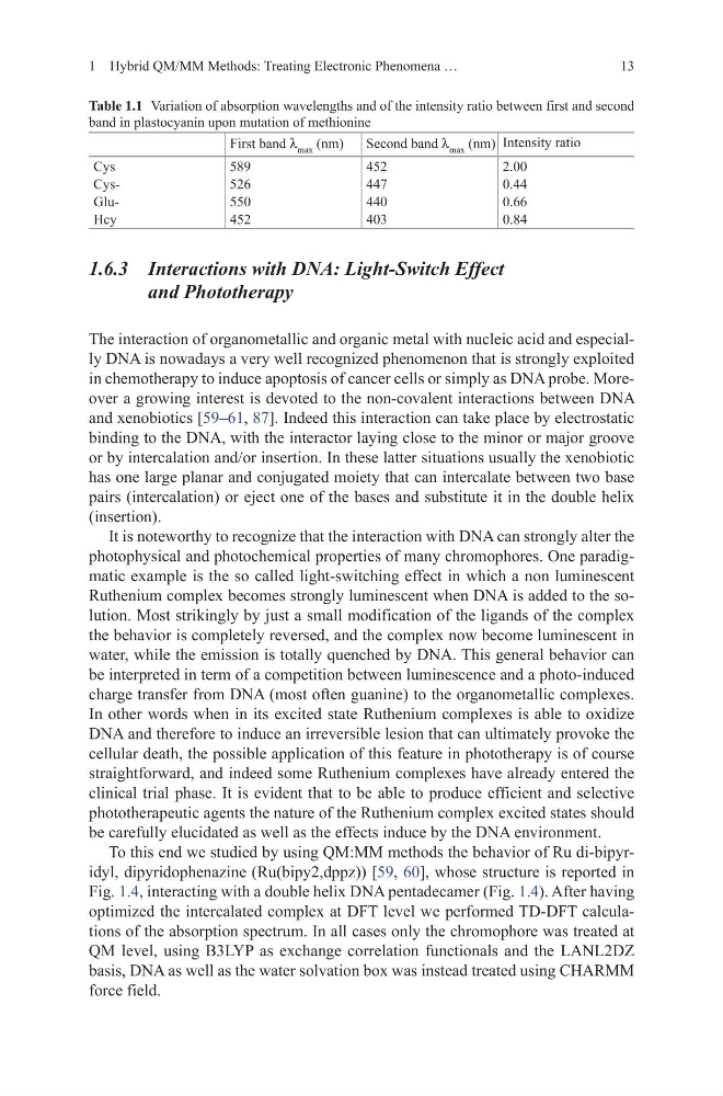
Table 1.1 Variation of absorption wavelengths and of the intensity ratio between first and second
band in plastocyanin upon mutation of methionine
First band λ max (nm)
Second band λ max (nm) Intensity ratio
Cys
589
452
2.00
Cys526
447
0.44
Glu550
440
0.66
Hcy
452
403
0.84
1 Hybrid QM/MM Methods: Treating Electronic Phenomena …
1.6.3 Interactions with DNA: Light-Switch Effect
and Phototherapy
The interaction of organometallic and organic metal with nucleic acid and especially DNA is nowadays a very well recognized phenomenon that is strongly exploited
in chemotherapy to induce apoptosis of cancer cells or simply as DNA probe. Moreover a growing interest is devoted to the non-covalent interactions between DNA
and xenobiotics [59–61, 87]. Indeed this interaction can take place by electrostatic
binding to the DNA, with the interactor laying close to the minor or major groove
or by intercalation and/or insertion. In these latter situations usually the xenobiotic
has one large planar and conjugated moiety that can intercalate between two base
pairs (intercalation) or eject one of the bases and substitute it in the double helix
(insertion).
It is noteworthy to recognize that the interaction with DNA can strongly alter the
photophysical and photochemical properties of many chromophores. One paradigmatic example is the so called light-switching effect in which a non luminescent
Ruthenium complex becomes strongly luminescent when DNA is added to the solution. Most strikingly by just a small modification of the ligands of the complex
the behavior is completely reversed, and the complex now become luminescent in
water, while the emission is totally quenched by DNA. This general behavior can
be interpreted in term of a competition between luminescence and a photo-induced
charge transfer from DNA (most often guanine) to the organometallic complexes.
In other words when in its excited state Ruthenium complexes is able to oxidize
DNA and therefore to induce an irreversible lesion that can ultimately provoke the
cellular death, the possible application of this feature in phototherapy is of course
straightforward, and indeed some Ruthenium complexes have already entered the
clinical trial phase. It is evident that to be able to produce efficient and selective
phototherapeutic agents the nature of the Ruthenium complex excited states should
be carefully elucidated as well as the effects induce by the DNA environment.
To this end we studied by using QM:MM methods the behavior of Ru di-bipyridyl, dipyridophenazine (Ru(bipy2,dppz)) [59, 60], whose structure is reported in
Fig. 1.4, interacting with a double helix DNA pentadecamer (Fig. 1.4). After having
optimized the intercalated complex at DFT level we performed TD-DFT calculations of the absorption spectrum. In all cases only the chromophore was treated at
QM level, using B3LYP as exchange correlation functionals and the LANL2DZ
basis, DNA as well as the water solvation box was instead treated using CHARMM
force field.
Table 1.1 Variation of absorption wavelengths and of the intensity ratio between first and second
band in plastocyanin upon mutation of methionine
First band λ max (nm)
Second band λ max (nm) Intensity ratio
Cys
589
452
2.00
Cys526
447
0.44
Glu550
440
0.66
Hcy
452
403
0.84