

223
basis sets were used for all the other atoms in the high layer. The ionized species
originated by removal of proton from the acidic functional group of ACE inhibitor.
Complexes containing ionized (non-protonated) ACE inhibitors are marked with
‘‘a” and complexes containing neutral (protonated) ACE inhibitors are marked with
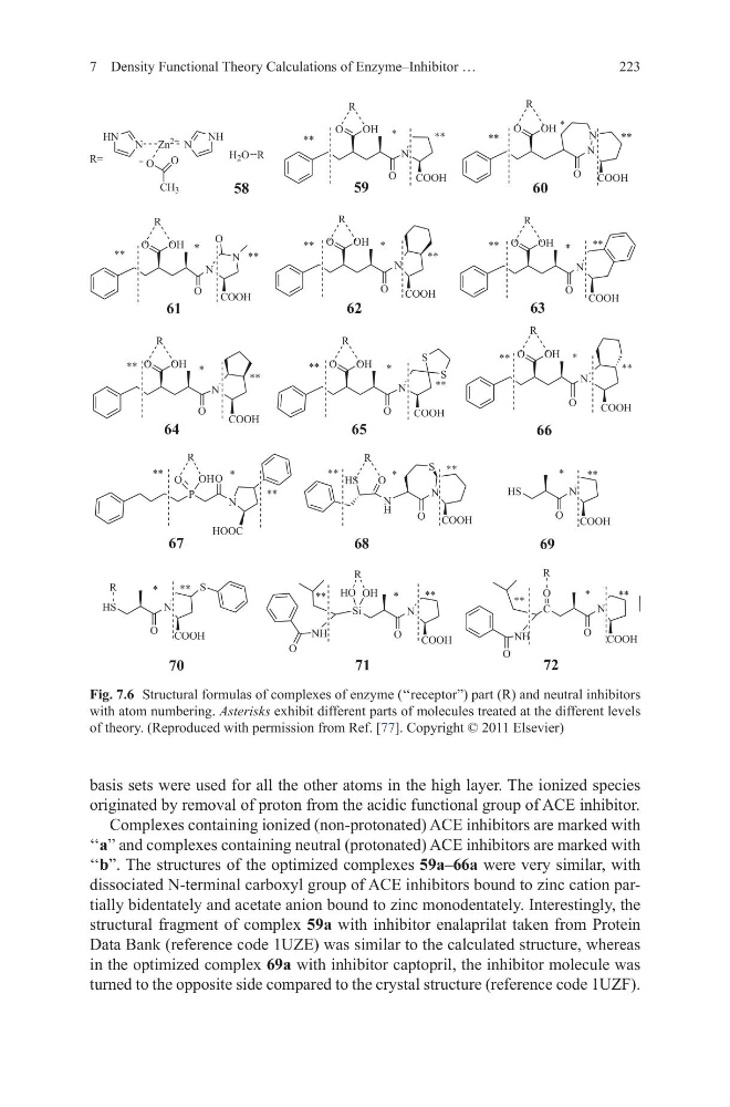
‘‘b”. The structures of the optimized complexes 59a–66a were very similar, with
dissociated N-terminal carboxyl group of ACE inhibitors bound to zinc cation partially bidentately and acetate anion bound to zinc monodentately. Interestingly, the
structural fragment of complex 59a with inhibitor enalaprilat taken from Protein
Data Bank (reference code 1UZE) was similar to the calculated structure, whereas
in the optimized complex 69a with inhibitor captopril, the inhibitor molecule was
turned to the opposite side compared to the crystal structure (reference code 1UZF).
7 Density Functional Theory Calculations of Enzyme–Inhibitor …
+ 2 5
2
1
2+
2
&22+
2
1
1
2+
2
&22+
5
5
2
1
1
2+
2
&22+
2
5
2
1
2+
2
&22+
2
1
2+
2
&22+
5
5
2
1
2+
2
&22+
2
1
2+
2
&22+
6
6
5
5
2
1
2+
2
&22+
5
3
1
2
+22&
1
+
2
1
6
&22+
+6
+6
2
1
&22+
2+
2
5
2
5
+6
2
1
&22+
6
6L
2
1
&22+
1+
2
2+
+2
5
5
&
2
1
&22+
1+
2
2
5
+1 1 =Q
1+
1
2
2
&+
5
Fig. 7.6 Structural formulas of complexes of enzyme (‘‘receptor”) part (R) and neutral inhibitors
with atom numbering. Asterisks exhibit different parts of molecules treated at the different levels
of theory. (Reproduced with permission from Ref. [77]. Copyright © 2011 Elsevier)
basis sets were used for all the other atoms in the high layer. The ionized species
originated by removal of proton from the acidic functional group of ACE inhibitor.
Complexes containing ionized (non-protonated) ACE inhibitors are marked with
‘‘a” and complexes containing neutral (protonated) ACE inhibitors are marked with
‘‘b”. The structures of the optimized complexes 59a–66a were very similar, with
dissociated N-terminal carboxyl group of ACE inhibitors bound to zinc cation partially bidentately and acetate anion bound to zinc monodentately. Interestingly, the
structural fragment of complex 59a with inhibitor enalaprilat taken from Protein
Data Bank (reference code 1UZE) was similar to the calculated structure, whereas
in the optimized complex 69a with inhibitor captopril, the inhibitor molecule was
turned to the opposite side compared to the crystal structure (reference code 1UZF).
7 Density Functional Theory Calculations of Enzyme–Inhibitor …
+ 2 5
2
1
2+
2
&22+
2
1
1
2+
2
&22+
5
5
2
1
1
2+
2
&22+
2
5
2
1
2+
2
&22+
2
1
2+
2
&22+
5
5
2
1
2+
2
&22+
2
1
2+
2
&22+
6
6
5
5
2
1
2+
2
&22+
5
3
1
2
+22&
1
+
2
1
6
&22+
+6
+6
2
1
&22+
2+
2
5
2
5
+6
2
1
&22+
6
6L
2
1
&22+
1+
2
2+
+2
5
5
&
2
1
&22+
1+
2
2
5
+1 1 =Q
1+
1
2
2
&+
5
Fig. 7.6 Structural formulas of complexes of enzyme (‘‘receptor”) part (R) and neutral inhibitors
with atom numbering. Asterisks exhibit different parts of molecules treated at the different levels
of theory. (Reproduced with permission from Ref. [77]. Copyright © 2011 Elsevier)