

6. Vacuum coating equipment for metallization: Explorer DualChamber, Denton Vacuum, USA (see Note 4).
7. High frequency pulse power supply: DuPR10-1-3, Dynatronix, USA (see Note 5).
8. Platinum-plated titanium anode.
9. Magnetic hot plate stirrers.
10. Microscope.
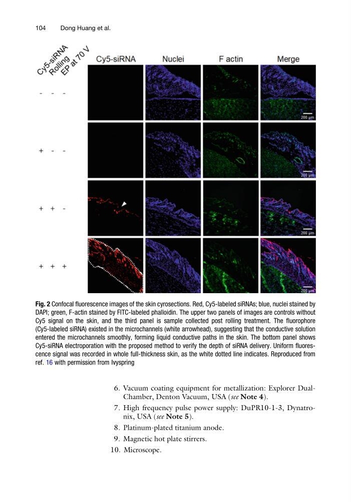
Fig. 2 Confocal fluorescence images of the skin cyrosections. Red, Cy5-labeled siRNAs; blue, nuclei stained by
DAPI; green, F-actin stained by FITC-labeled phalloidin. The upper two panels of images are controls without
Cy5 signal on the skin, and the third panel is sample collected post rolling treatment. The fluorophore
(Cy5-labeled siRNA) existed in the microchannels (white arrowhead), suggesting that the conductive solution
entered the microchannels smoothly, forming liquid conductive paths in the skin. The bottom panel shows
Cy5-siRNA electroporation with the proposed method to verify the depth of siRNA delivery. Uniform fluorescence signal was recorded in whole full-thickness skin, as the white dotted line indicates. Reproduced from
ref. 16 with permission from Ivyspring
104
Dong Huang et al.
7. High frequency pulse power supply: DuPR10-1-3, Dynatronix, USA (see Note 5).
8. Platinum-plated titanium anode.
9. Magnetic hot plate stirrers.
10. Microscope.
Fig. 2 Confocal fluorescence images of the skin cyrosections. Red, Cy5-labeled siRNAs; blue, nuclei stained by
DAPI; green, F-actin stained by FITC-labeled phalloidin. The upper two panels of images are controls without
Cy5 signal on the skin, and the third panel is sample collected post rolling treatment. The fluorophore
(Cy5-labeled siRNA) existed in the microchannels (white arrowhead), suggesting that the conductive solution
entered the microchannels smoothly, forming liquid conductive paths in the skin. The bottom panel shows
Cy5-siRNA electroporation with the proposed method to verify the depth of siRNA delivery. Uniform fluorescence signal was recorded in whole full-thickness skin, as the white dotted line indicates. Reproduced from
ref. 16 with permission from Ivyspring
104
Dong Huang et al.