

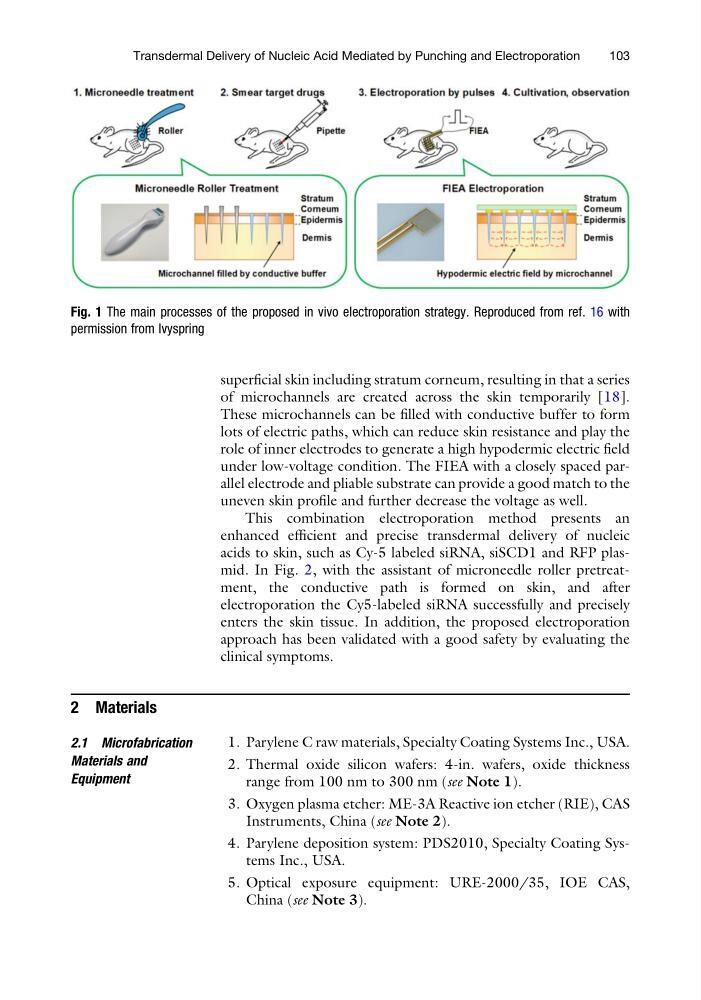
superficial skin including stratum corneum, resulting in that a series
of microchannels are created across the skin temporarily [18].
These microchannels can be filled with conductive buffer to form
lots of electric paths, which can reduce skin resistance and play the
role of inner electrodes to generate a high hypodermic electric field
under low-voltage condition. The FIEA with a closely spaced parallel electrode and pliable substrate can provide a good match to the
uneven skin profile and further decrease the voltage as well.
This combination electroporation method presents an
enhanced efficient and precise transdermal delivery of nucleic
acids to skin, such as Cy-5 labeled siRNA, siSCD1 and RFP plasmid. In Fig. 2, with the assistant of microneedle roller pretreatment, the conductive path is formed on skin, and after
electroporation the Cy5-labeled siRNA successfully and precisely
enters the skin tissue. In addition, the proposed electroporation
approach has been validated with a good safety by evaluating the
clinical symptoms.
2 Materials
2.1 Microfabrication
Materials and
Equipment
1. Parylene C raw materials, Specialty Coating Systems Inc., USA.
2. Thermal oxide silicon wafers: 4-in. wafers, oxide thickness
range from 100 nm to 300 nm (see Note 1).
3. Oxygen plasma etcher: ME-3A Reactive ion etcher (RIE), CAS
Instruments, China (see Note 2).
4. Parylene deposition system: PDS2010, Specialty Coating Systems Inc., USA.
5. Optical exposure equipment: URE-2000/35, IOE CAS,
China (see Note 3).
Fig. 1 The main processes of the proposed in vivo electroporation strategy. Reproduced from ref. 16 with
permission from Ivyspring
Transdermal Delivery of Nucleic Acid Mediated by Punching and Electroporation
103
of microchannels are created across the skin temporarily [18].
These microchannels can be filled with conductive buffer to form
lots of electric paths, which can reduce skin resistance and play the
role of inner electrodes to generate a high hypodermic electric field
under low-voltage condition. The FIEA with a closely spaced parallel electrode and pliable substrate can provide a good match to the
uneven skin profile and further decrease the voltage as well.
This combination electroporation method presents an
enhanced efficient and precise transdermal delivery of nucleic
acids to skin, such as Cy-5 labeled siRNA, siSCD1 and RFP plasmid. In Fig. 2, with the assistant of microneedle roller pretreatment, the conductive path is formed on skin, and after
electroporation the Cy5-labeled siRNA successfully and precisely
enters the skin tissue. In addition, the proposed electroporation
approach has been validated with a good safety by evaluating the
clinical symptoms.
2 Materials
2.1 Microfabrication
Materials and
Equipment
1. Parylene C raw materials, Specialty Coating Systems Inc., USA.
2. Thermal oxide silicon wafers: 4-in. wafers, oxide thickness
range from 100 nm to 300 nm (see Note 1).
3. Oxygen plasma etcher: ME-3A Reactive ion etcher (RIE), CAS
Instruments, China (see Note 2).
4. Parylene deposition system: PDS2010, Specialty Coating Systems Inc., USA.
5. Optical exposure equipment: URE-2000/35, IOE CAS,
China (see Note 3).
Fig. 1 The main processes of the proposed in vivo electroporation strategy. Reproduced from ref. 16 with
permission from Ivyspring
Transdermal Delivery of Nucleic Acid Mediated by Punching and Electroporation
103