

2 XUV Lasers for Ultrafast Electronic Control in H 2
35
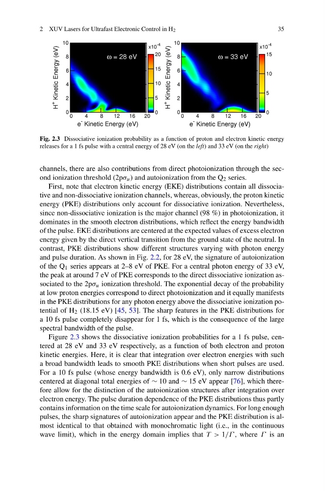
Fig. 2.3 Dissociative ionization probability as a function of proton and electron kinetic energy
releases for a 1 fs pulse with a central energy of 28 eV (on the left) and 33 eV (on the right)
channels, there are also contributions from direct photoionization through the second ionization threshold (2pσ u ) and autoionization from the Q 2 series.
First, note that electron kinetic energy (EKE) distributions contain all dissociative and non-dissociative ionization channels, whereas, obviously, the proton kinetic
energy (PKE) distributions only account for dissociative ionization. Nevertheless,
since non-dissociative ionization is the major channel (98 %) in photoionization, it
dominates in the smooth electron distributions, which reflect the energy bandwidth
of the pulse. EKE distributions are centered at the expected values of excess electron
energy given by the direct vertical transition from the ground state of the neutral. In
contrast, PKE distributions show different structures varying with photon energy
and pulse duration. As shown in Fig. 2.2, for 28 eV, the signature of autoionization
of the Q 1 series appears at 2–8 eV of PKE. For a central photon energy of 33 eV,
the peak at around 7 eV of PKE corresponds to the direct dissociative ionization associated to the 2pσ u ionization threshold. The exponential decay of the probability
at low proton energies correspond to direct photoionization and it equally manifests
in the PKE distributions for any photon energy above the dissociative ionization potential of H 2 (18.15 eV) [45, 53]. The sharp features in the PKE distributions for
a 10 fs pulse completely disappear for 1 fs, which is the consequence of the large
spectral bandwidth of the pulse.
Figure 2.3 shows the dissociative ionization probabilities for a 1 fs pulse, centered at 28 eV and 33 eV respectively, as a function of both electron and proton
kinetic energies. Here, it is clear that integration over electron energies with such
a broad bandwidth leads to smooth PKE distributions when short pulses are used.
For a 10 fs pulse (whose energy bandwidth is 0.6 eV), only narrow distributions
centered at diagonal total energies of ∼ 10 and ∼ 15 eV appear [76], which therefore allow for the distinction of the autoionization structures after integration over
electron energy. The pulse duration dependence of the PKE distributions thus partly
contains information on the time scale for autoionization dynamics. For long enough
pulses, the sharp signatures of autoionization appear and the PKE distribution is almost identical to that obtained with monochromatic light (i.e., in the continuous
wave limit), which in the energy domain implies that T > 1/Γ , where Γ is an
35
Fig. 2.3 Dissociative ionization probability as a function of proton and electron kinetic energy
releases for a 1 fs pulse with a central energy of 28 eV (on the left) and 33 eV (on the right)
channels, there are also contributions from direct photoionization through the second ionization threshold (2pσ u ) and autoionization from the Q 2 series.
First, note that electron kinetic energy (EKE) distributions contain all dissociative and non-dissociative ionization channels, whereas, obviously, the proton kinetic
energy (PKE) distributions only account for dissociative ionization. Nevertheless,
since non-dissociative ionization is the major channel (98 %) in photoionization, it
dominates in the smooth electron distributions, which reflect the energy bandwidth
of the pulse. EKE distributions are centered at the expected values of excess electron
energy given by the direct vertical transition from the ground state of the neutral. In
contrast, PKE distributions show different structures varying with photon energy
and pulse duration. As shown in Fig. 2.2, for 28 eV, the signature of autoionization
of the Q 1 series appears at 2–8 eV of PKE. For a central photon energy of 33 eV,
the peak at around 7 eV of PKE corresponds to the direct dissociative ionization associated to the 2pσ u ionization threshold. The exponential decay of the probability
at low proton energies correspond to direct photoionization and it equally manifests
in the PKE distributions for any photon energy above the dissociative ionization potential of H 2 (18.15 eV) [45, 53]. The sharp features in the PKE distributions for
a 10 fs pulse completely disappear for 1 fs, which is the consequence of the large
spectral bandwidth of the pulse.
Figure 2.3 shows the dissociative ionization probabilities for a 1 fs pulse, centered at 28 eV and 33 eV respectively, as a function of both electron and proton
kinetic energies. Here, it is clear that integration over electron energies with such
a broad bandwidth leads to smooth PKE distributions when short pulses are used.
For a 10 fs pulse (whose energy bandwidth is 0.6 eV), only narrow distributions
centered at diagonal total energies of ∼ 10 and ∼ 15 eV appear [76], which therefore allow for the distinction of the autoionization structures after integration over
electron energy. The pulse duration dependence of the PKE distributions thus partly
contains information on the time scale for autoionization dynamics. For long enough
pulses, the sharp signatures of autoionization appear and the PKE distribution is almost identical to that obtained with monochromatic light (i.e., in the continuous
wave limit), which in the energy domain implies that T > 1/Γ , where Γ is an