

8 Ultrafast Ionization and Fragmentation: From Small Molecules
191
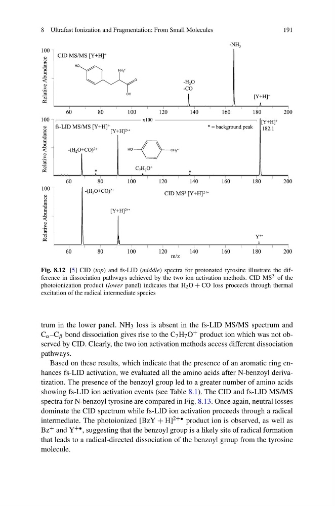
Fig. 8.12 [5] CID (top) and fs-LID (middle) spectra for protonated tyrosine illustrate the difference in dissociation pathways achieved by the two ion activation methods. CID MS 3 of the
photoionization product (lower panel) indicates that H 2 O + CO loss proceeds through thermal
excitation of the radical intermediate species
trum in the lower panel. NH 3 loss is absent in the fs-LID MS/MS spectrum and
C α –C β bond dissociation gives rise to the C 7 H 7 O + product ion which was not observed by CID. Clearly, the two ion activation methods access different dissociation
pathways.
Based on these results, which indicate that the presence of an aromatic ring enhances fs-LID activation, we evaluated all the amino acids after N-benzoyl derivatization. The presence of the benzoyl group led to a greater number of amino acids
showing fs-LID ion activation events (see Table 8.1). The CID and fs-LID MS/MS
spectra for N-benzoyl tyrosine are compared in Fig. 8.13. Once again, neutral losses
dominate the CID spectrum while fs-LID ion activation proceeds through a radical
intermediate. The photoionized [BzY + H] 2+• product ion is observed, as well as
Bz + and Y +• , suggesting that the benzoyl group is a likely site of radical formation
that leads to a radical-directed dissociation of the benzoyl group from the tyrosine
molecule.
191
Fig. 8.12 [5] CID (top) and fs-LID (middle) spectra for protonated tyrosine illustrate the difference in dissociation pathways achieved by the two ion activation methods. CID MS 3 of the
photoionization product (lower panel) indicates that H 2 O + CO loss proceeds through thermal
excitation of the radical intermediate species
trum in the lower panel. NH 3 loss is absent in the fs-LID MS/MS spectrum and
C α –C β bond dissociation gives rise to the C 7 H 7 O + product ion which was not observed by CID. Clearly, the two ion activation methods access different dissociation
pathways.
Based on these results, which indicate that the presence of an aromatic ring enhances fs-LID activation, we evaluated all the amino acids after N-benzoyl derivatization. The presence of the benzoyl group led to a greater number of amino acids
showing fs-LID ion activation events (see Table 8.1). The CID and fs-LID MS/MS
spectra for N-benzoyl tyrosine are compared in Fig. 8.13. Once again, neutral losses
dominate the CID spectrum while fs-LID ion activation proceeds through a radical
intermediate. The photoionized [BzY + H] 2+• product ion is observed, as well as
Bz + and Y +• , suggesting that the benzoyl group is a likely site of radical formation
that leads to a radical-directed dissociation of the benzoyl group from the tyrosine
molecule.