

the donation of an unshared electron pair. In the case of ester-based donors, the
coordination accompanies a red-shift of the C¼O vibrational frequency, whose
degree depends on the chemical nature of the Lewis acidic sites. This fact has been
frequently utilized to examine the state and location of ester-type donors in solid
catalysts by means of IR spectroscopy. Terano et al. clarified with IR spectroscopy
and thermal analysis that EB dominantly resides on MgCl 2 surfaces without
forming a TiCl 4 ·EB complex [30]. This is in agreement with quantum chemical
calculations, which concluded that the dissociative adsorption of TiCl 4 ·EB on
MgCl 2 surfaces is energetically more advantageous than non-dissociative adsorption [31]. Similar IR results were obtained for dibutylphthalate (DBP), for example,
by Arzoumanidis and Karayannis [32] They studied catalysts that were activated at
different temperatures and found that DBP dominantly coordinated to surface Mg
sites at any activation temperature. However, a residual amount of TiCl 4 ·DBP
complex was detected only when a catalyst was activated at a too-low temperature
(called under-activation), whereas activation at a too-high temperature led to the
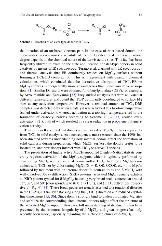
formation of carbonyl halides according to Scheme 1 [32, 33] (called overactivation [32]), both of which resulted in a clear reduction in propylene polymerization activity.
Thus, it is well accepted that donors are supported on MgCl 2 surfaces separately
from TiCl 4 in solid catalysts. As a consequence, most research since the 1990s has
been directed towards understanding how internal donors affect the formation of
solid catalysts during preparation, which MgCl 2 surfaces the donors prefer to be
located on, and how donors interact with TiCl 4 or active Ti species.
The preparation of highly active MgCl 2 -supported Ziegler–Natta catalysts generally requires activation of the MgCl 2 support, which is typically performed by
co-grinding MgCl 2 with an internal donor and/or TiCl 4 , treating a MgCl 2 ·donor
adduct with TiCl 4 , or by chlorinating MgX 2 (X ¼ R, OR, OCOR, etc.) into MgCl 2
followed by treatment with an internal donor. In contrast to α- and β-MgCl 2 with
well-dissolved X-ray diffraction (XRD) patterns, activated MgCl 2 usually exhibits
an XRD pattern typical for δ-MgCl 2 , featuring very broad peaks centered at around
15
, 32
, and 50
[corresponding to (0 0 3), (1 0 l), and (1 1 0) reflections, respectively] (Fig. 6) [34]. These broad peaks are usually ascribed to a rotational disorder
in the Cl–Mg–Cl tri-layer stacking along the (0 0 1) direction and reduced crystalline dimensions [35, 36]. Since donors strongly bind to undercoordinated Mg sites
and stabilize the corresponding sites, internal donors might affect the structure of
the activated MgCl 2 support. However, full understanding of its structure has been
prevented by the structural irregularity of δ-MgCl 2 , and great progress has only
recently been made, especially regarding the surface structures of δ-MgCl 2 .
Scheme 1 Reaction of an ester-type donor with TiCl 4
The Use of Donors to Increase the Isotacticity of Polypropylene
87
coordination accompanies a red-shift of the C¼O vibrational frequency, whose
degree depends on the chemical nature of the Lewis acidic sites. This fact has been
frequently utilized to examine the state and location of ester-type donors in solid
catalysts by means of IR spectroscopy. Terano et al. clarified with IR spectroscopy
and thermal analysis that EB dominantly resides on MgCl 2 surfaces without
forming a TiCl 4 ·EB complex [30]. This is in agreement with quantum chemical
calculations, which concluded that the dissociative adsorption of TiCl 4 ·EB on
MgCl 2 surfaces is energetically more advantageous than non-dissociative adsorption [31]. Similar IR results were obtained for dibutylphthalate (DBP), for example,
by Arzoumanidis and Karayannis [32] They studied catalysts that were activated at
different temperatures and found that DBP dominantly coordinated to surface Mg
sites at any activation temperature. However, a residual amount of TiCl 4 ·DBP
complex was detected only when a catalyst was activated at a too-low temperature
(called under-activation), whereas activation at a too-high temperature led to the
formation of carbonyl halides according to Scheme 1 [32, 33] (called overactivation [32]), both of which resulted in a clear reduction in propylene polymerization activity.
Thus, it is well accepted that donors are supported on MgCl 2 surfaces separately
from TiCl 4 in solid catalysts. As a consequence, most research since the 1990s has
been directed towards understanding how internal donors affect the formation of
solid catalysts during preparation, which MgCl 2 surfaces the donors prefer to be
located on, and how donors interact with TiCl 4 or active Ti species.
The preparation of highly active MgCl 2 -supported Ziegler–Natta catalysts generally requires activation of the MgCl 2 support, which is typically performed by
co-grinding MgCl 2 with an internal donor and/or TiCl 4 , treating a MgCl 2 ·donor
adduct with TiCl 4 , or by chlorinating MgX 2 (X ¼ R, OR, OCOR, etc.) into MgCl 2
followed by treatment with an internal donor. In contrast to α- and β-MgCl 2 with
well-dissolved X-ray diffraction (XRD) patterns, activated MgCl 2 usually exhibits
an XRD pattern typical for δ-MgCl 2 , featuring very broad peaks centered at around
15
, 32
, and 50
[corresponding to (0 0 3), (1 0 l), and (1 1 0) reflections, respectively] (Fig. 6) [34]. These broad peaks are usually ascribed to a rotational disorder
in the Cl–Mg–Cl tri-layer stacking along the (0 0 1) direction and reduced crystalline dimensions [35, 36]. Since donors strongly bind to undercoordinated Mg sites
and stabilize the corresponding sites, internal donors might affect the structure of
the activated MgCl 2 support. However, full understanding of its structure has been
prevented by the structural irregularity of δ-MgCl 2 , and great progress has only
recently been made, especially regarding the surface structures of δ-MgCl 2 .
Scheme 1 Reaction of an ester-type donor with TiCl 4
The Use of Donors to Increase the Isotacticity of Polypropylene
87