

358
L. Pan and X. Chen
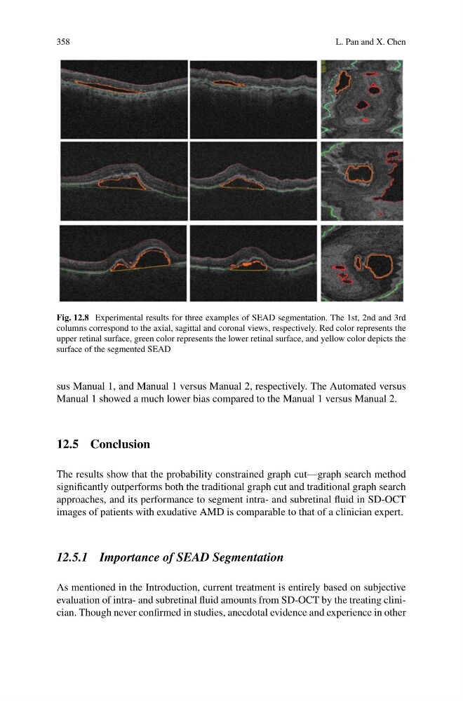
Fig. 12.8 Experimental results for three examples of SEAD segmentation. The 1st, 2nd and 3rd
columns correspond to the axial, sagittal and coronal views, respectively. Red color represents the
upper retinal surface, green color represents the lower retinal surface, and yellow color depicts the
surface of the segmented SEAD
sus Manual 1, and Manual 1 versus Manual 2, respectively. The Automated versus
Manual 1 showed a much lower bias compared to the Manual 1 versus Manual 2.
12.5 Conclusion
The results show that the probability constrained graph cut—graph search method
significantly outperforms both the traditional graph cut and traditional graph search
approaches, and its performance to segment intra- and subretinal fluid in SD-OCT
images of patients with exudative AMD is comparable to that of a clinician expert.
12.5.1 Importance of SEAD Segmentation
As mentioned in the Introduction, current treatment is entirely based on subjective
evaluation of intra- and subretinal fluid amounts from SD-OCT by the treating clinician. Though never confirmed in studies, anecdotal evidence and experience in other
L. Pan and X. Chen
Fig. 12.8 Experimental results for three examples of SEAD segmentation. The 1st, 2nd and 3rd
columns correspond to the axial, sagittal and coronal views, respectively. Red color represents the
upper retinal surface, green color represents the lower retinal surface, and yellow color depicts the
surface of the segmented SEAD
sus Manual 1, and Manual 1 versus Manual 2, respectively. The Automated versus
Manual 1 showed a much lower bias compared to the Manual 1 versus Manual 2.
12.5 Conclusion
The results show that the probability constrained graph cut—graph search method
significantly outperforms both the traditional graph cut and traditional graph search
approaches, and its performance to segment intra- and subretinal fluid in SD-OCT
images of patients with exudative AMD is comparable to that of a clinician expert.
12.5.1 Importance of SEAD Segmentation
As mentioned in the Introduction, current treatment is entirely based on subjective
evaluation of intra- and subretinal fluid amounts from SD-OCT by the treating clinician. Though never confirmed in studies, anecdotal evidence and experience in other