

11 Segmentation and Visualization of Drusen …
327
(a)
(b)
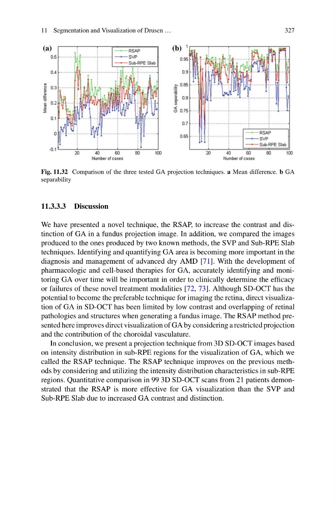
Fig. 11.32 Comparison of the three tested GA projection techniques. a Mean difference. b GA
separability
11.3.3.3 Discussion
We have presented a novel technique, the RSAP, to increase the contrast and distinction of GA in a fundus projection image. In addition, we compared the images
produced to the ones produced by two known methods, the SVP and Sub-RPE Slab
techniques. Identifying and quantifying GA area is becoming more important in the
diagnosis and management of advanced dry AMD [71]. With the development of
pharmacologic and cell-based therapies for GA, accurately identifying and monitoring GA over time will be important in order to clinically determine the efficacy
or failures of these novel treatment modalities [72, 73]. Although SD-OCT has the
potential to become the preferable technique for imaging the retina, direct visualization of GA in SD-OCT has been limited by low contrast and overlapping of retinal
pathologies and structures when generating a fundus image. The RSAP method presented here improves direct visualization of GA by considering a restricted projection
and the contribution of the choroidal vasculature.
In conclusion, we present a projection technique from 3D SD-OCT images based
on intensity distribution in sub-RPE regions for the visualization of GA, which we
called the RSAP technique. The RSAP technique improves on the previous methods by considering and utilizing the intensity distribution characteristics in sub-RPE
regions. Quantitative comparison in 99 3D SD-OCT scans from 21 patients demonstrated that the RSAP is more effective for GA visualization than the SVP and
Sub-RPE Slab due to increased GA contrast and distinction.
327
(a)
(b)
Fig. 11.32 Comparison of the three tested GA projection techniques. a Mean difference. b GA
separability
11.3.3.3 Discussion
We have presented a novel technique, the RSAP, to increase the contrast and distinction of GA in a fundus projection image. In addition, we compared the images
produced to the ones produced by two known methods, the SVP and Sub-RPE Slab
techniques. Identifying and quantifying GA area is becoming more important in the
diagnosis and management of advanced dry AMD [71]. With the development of
pharmacologic and cell-based therapies for GA, accurately identifying and monitoring GA over time will be important in order to clinically determine the efficacy
or failures of these novel treatment modalities [72, 73]. Although SD-OCT has the
potential to become the preferable technique for imaging the retina, direct visualization of GA in SD-OCT has been limited by low contrast and overlapping of retinal
pathologies and structures when generating a fundus image. The RSAP method presented here improves direct visualization of GA by considering a restricted projection
and the contribution of the choroidal vasculature.
In conclusion, we present a projection technique from 3D SD-OCT images based
on intensity distribution in sub-RPE regions for the visualization of GA, which we
called the RSAP technique. The RSAP technique improves on the previous methods by considering and utilizing the intensity distribution characteristics in sub-RPE
regions. Quantitative comparison in 99 3D SD-OCT scans from 21 patients demonstrated that the RSAP is more effective for GA visualization than the SVP and
Sub-RPE Slab due to increased GA contrast and distinction.