

328
Q. Chen et al.
(a)
(d)
(b)
(c)
GA
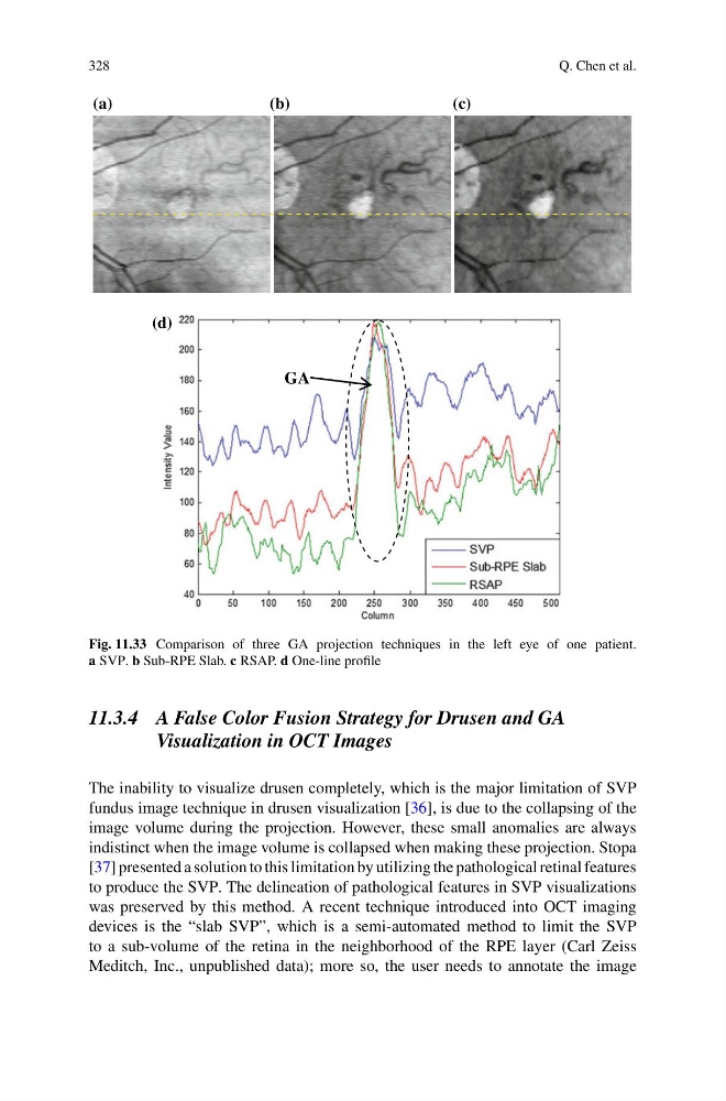
Fig. 11.33 Comparison of three GA projection techniques in the left eye of one patient.
a SVP. b Sub-RPE Slab. c RSAP. d One-line profile
11.3.4 A False Color Fusion Strategy for Drusen and GA
Visualization in OCT Images
The inability to visualize drusen completely, which is the major limitation of SVP
fundus image technique in drusen visualization [36], is due to the collapsing of the
image volume during the projection. However, these small anomalies are always
indistinct when the image volume is collapsed when making these projection. Stopa
[37] presented a solution to this limitation by utilizing the pathological retinal features
to produce the SVP. The delineation of pathological features in SVP visualizations
was preserved by this method. A recent technique introduced into OCT imaging
devices is the “slab SVP”, which is a semi-automated method to limit the SVP
to a sub-volume of the retina in the neighborhood of the RPE layer (Carl Zeiss
Meditch, Inc., unpublished data); more so, the user needs to annotate the image
Q. Chen et al.
(a)
(d)
(b)
(c)
GA
Fig. 11.33 Comparison of three GA projection techniques in the left eye of one patient.
a SVP. b Sub-RPE Slab. c RSAP. d One-line profile
11.3.4 A False Color Fusion Strategy for Drusen and GA
Visualization in OCT Images
The inability to visualize drusen completely, which is the major limitation of SVP
fundus image technique in drusen visualization [36], is due to the collapsing of the
image volume during the projection. However, these small anomalies are always
indistinct when the image volume is collapsed when making these projection. Stopa
[37] presented a solution to this limitation by utilizing the pathological retinal features
to produce the SVP. The delineation of pathological features in SVP visualizations
was preserved by this method. A recent technique introduced into OCT imaging
devices is the “slab SVP”, which is a semi-automated method to limit the SVP
to a sub-volume of the retina in the neighborhood of the RPE layer (Carl Zeiss
Meditch, Inc., unpublished data); more so, the user needs to annotate the image