

1 Clinical Applications of Retinal Optical Coherence Tomography
19
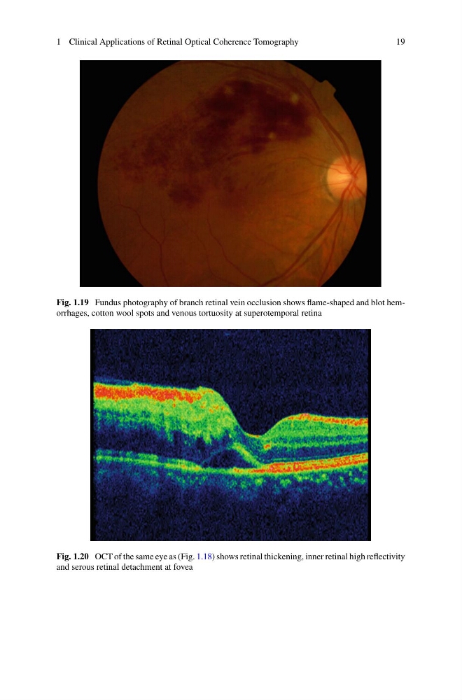
Fig. 1.19 Fundus photography of branch retinal vein occlusion shows flame-shaped and blot hemorrhages, cotton wool spots and venous tortuosity at superotemporal retina
Fig. 1.20 OCT of the same eye as (Fig. 1.18) shows retinal thickening, inner retinal high reflectivity
and serous retinal detachment at fovea
19
Fig. 1.19 Fundus photography of branch retinal vein occlusion shows flame-shaped and blot hemorrhages, cotton wool spots and venous tortuosity at superotemporal retina
Fig. 1.20 OCT of the same eye as (Fig. 1.18) shows retinal thickening, inner retinal high reflectivity
and serous retinal detachment at fovea