

20
H. Chen et al.
Drusen is degenerative nodular formations located in Bruch’s membrane and
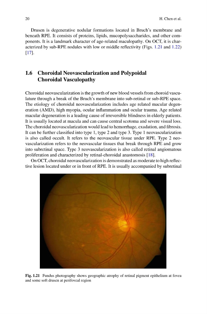
beneath RPE. It consists of proteins, lipids, mucopolysaccharides, and other components. It is a landmark character of age-related maculopathy. On OCT, it is characterized by sub-RPE nodules with low or middle reflectivity (Figs. 1.21 and 1.22)
[17].
1.6 Choroidal Neovascularization and Polypoidal
Choroidal Vasculopathy
Choroidal neovascularization is the growth of new blood vessels from choroid vasculature through a break of the Bruch’s membrane into sub-retinal or sub-RPE space.
The etiology of choroidal neovascularization includes age related macular degeneration (AMD), high myopia, ocular inflammation and ocular trauma. Age related
macular degeneration is a leading cause of irreversible blindness in elderly patients.
It is usually located at macula and can cause central scotoma and severe visual loss.
The choroidal neovascularization would lead to hemorrhage, exudation, and fibrosis.
It can be further classified into type 1, type 2 and type 3. Type 1 neovascularization
is also called occult. It refers to the neovascular tissue under RPE. Type 2 neovascularization refers to the neovascular tissues that break through RPE and grow
into subretinal space. Type 3 neovascularization is also called retinal angiomatous
proliferation and characterized by retinal-choroidal anastomosis [18].
On OCT, choroidal neovascularization is demonstrated as moderate to high reflective lesion located under or in front of RPE. It is usually accompanied by subretinal
Fig. 1.21 Fundus photography shows geographic atrophy of retinal pigment epithelium at fovea
and some soft drusen at perifoveal region
H. Chen et al.
Drusen is degenerative nodular formations located in Bruch’s membrane and
beneath RPE. It consists of proteins, lipids, mucopolysaccharides, and other components. It is a landmark character of age-related maculopathy. On OCT, it is characterized by sub-RPE nodules with low or middle reflectivity (Figs. 1.21 and 1.22)
[17].
1.6 Choroidal Neovascularization and Polypoidal
Choroidal Vasculopathy
Choroidal neovascularization is the growth of new blood vessels from choroid vasculature through a break of the Bruch’s membrane into sub-retinal or sub-RPE space.
The etiology of choroidal neovascularization includes age related macular degeneration (AMD), high myopia, ocular inflammation and ocular trauma. Age related
macular degeneration is a leading cause of irreversible blindness in elderly patients.
It is usually located at macula and can cause central scotoma and severe visual loss.
The choroidal neovascularization would lead to hemorrhage, exudation, and fibrosis.
It can be further classified into type 1, type 2 and type 3. Type 1 neovascularization
is also called occult. It refers to the neovascular tissue under RPE. Type 2 neovascularization refers to the neovascular tissues that break through RPE and grow
into subretinal space. Type 3 neovascularization is also called retinal angiomatous
proliferation and characterized by retinal-choroidal anastomosis [18].
On OCT, choroidal neovascularization is demonstrated as moderate to high reflective lesion located under or in front of RPE. It is usually accompanied by subretinal
Fig. 1.21 Fundus photography shows geographic atrophy of retinal pigment epithelium at fovea
and some soft drusen at perifoveal region