

18
H. Chen et al.
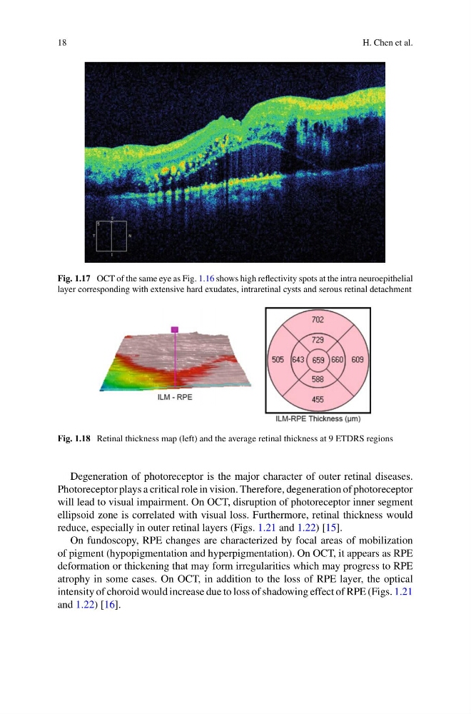
Fig. 1.17 OCT of the same eye as Fig. 1.16 shows high reflectivity spots at the intra neuroepithelial
layer corresponding with extensive hard exudates, intraretinal cysts and serous retinal detachment
Fig. 1.18 Retinal thickness map (left) and the average retinal thickness at 9 ETDRS regions
Degeneration of photoreceptor is the major character of outer retinal diseases.
Photoreceptor plays a critical role in vision. Therefore, degeneration of photoreceptor
will lead to visual impairment. On OCT, disruption of photoreceptor inner segment
ellipsoid zone is correlated with visual loss. Furthermore, retinal thickness would
reduce, especially in outer retinal layers (Figs. 1.21 and 1.22) [15].
On fundoscopy, RPE changes are characterized by focal areas of mobilization
of pigment (hypopigmentation and hyperpigmentation). On OCT, it appears as RPE
deformation or thickening that may form irregularities which may progress to RPE
atrophy in some cases. On OCT, in addition to the loss of RPE layer, the optical
intensity of choroid would increase due to loss of shadowing effect of RPE (Figs. 1.21
and 1.22) [16].
H. Chen et al.
Fig. 1.17 OCT of the same eye as Fig. 1.16 shows high reflectivity spots at the intra neuroepithelial
layer corresponding with extensive hard exudates, intraretinal cysts and serous retinal detachment
Fig. 1.18 Retinal thickness map (left) and the average retinal thickness at 9 ETDRS regions
Degeneration of photoreceptor is the major character of outer retinal diseases.
Photoreceptor plays a critical role in vision. Therefore, degeneration of photoreceptor
will lead to visual impairment. On OCT, disruption of photoreceptor inner segment
ellipsoid zone is correlated with visual loss. Furthermore, retinal thickness would
reduce, especially in outer retinal layers (Figs. 1.21 and 1.22) [15].
On fundoscopy, RPE changes are characterized by focal areas of mobilization
of pigment (hypopigmentation and hyperpigmentation). On OCT, it appears as RPE
deformation or thickening that may form irregularities which may progress to RPE
atrophy in some cases. On OCT, in addition to the loss of RPE layer, the optical
intensity of choroid would increase due to loss of shadowing effect of RPE (Figs. 1.21
and 1.22) [16].