

1 Clinical Applications of Retinal Optical Coherence Tomography
17
1.4.3 Retinal Vein Occlusion
Retinal vein occlusion may involve either the central or branch retinal veins. The fundal manifestation of retinal vein occlusion also includes retinal hemorrhage, cotton
wool spots, retinal hard exudates, macular edema. The OCT appearances are similar
to those of diabetic retinopathy (Figs. 1.19 and 1.20).
1.5 Outer Retinal Degenerative Diseases
Outer retinal layers include outer nuclear layer, photoreceptor inner and outer segment, retinal pigment epithelium. Degeneration of outer retina can be caused by
genetic mutation, or secondary to other factors, including traumatic impact, retinal
detachment, inflammation, toxicity, or age related macular degeneration.
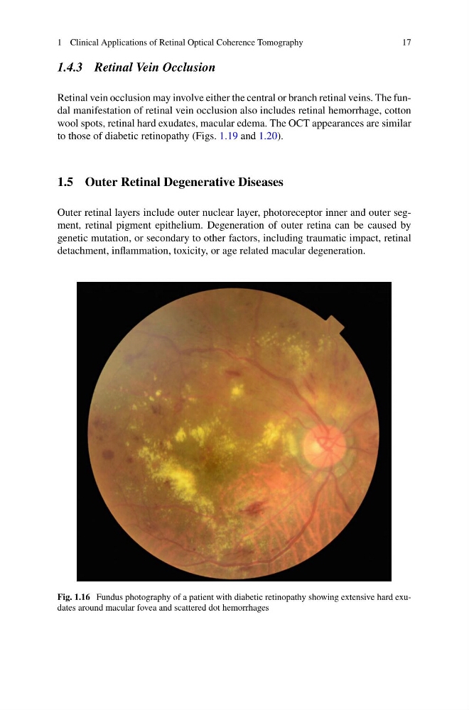
Fig. 1.16 Fundus photography of a patient with diabetic retinopathy showing extensive hard exudates around macular fovea and scattered dot hemorrhages
17
1.4.3 Retinal Vein Occlusion
Retinal vein occlusion may involve either the central or branch retinal veins. The fundal manifestation of retinal vein occlusion also includes retinal hemorrhage, cotton
wool spots, retinal hard exudates, macular edema. The OCT appearances are similar
to those of diabetic retinopathy (Figs. 1.19 and 1.20).
1.5 Outer Retinal Degenerative Diseases
Outer retinal layers include outer nuclear layer, photoreceptor inner and outer segment, retinal pigment epithelium. Degeneration of outer retina can be caused by
genetic mutation, or secondary to other factors, including traumatic impact, retinal
detachment, inflammation, toxicity, or age related macular degeneration.
Fig. 1.16 Fundus photography of a patient with diabetic retinopathy showing extensive hard exudates around macular fovea and scattered dot hemorrhages