

16
H. Chen et al.
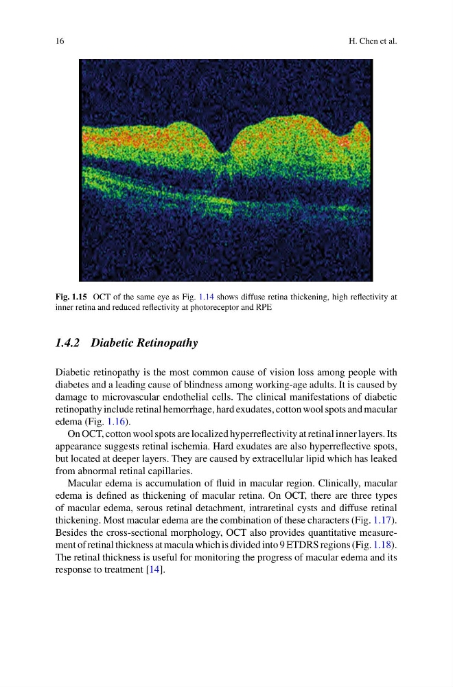
Fig. 1.15 OCT of the same eye as Fig. 1.14 shows diffuse retina thickening, high reflectivity at
inner retina and reduced reflectivity at photoreceptor and RPE
1.4.2 Diabetic Retinopathy
Diabetic retinopathy is the most common cause of vision loss among people with
diabetes and a leading cause of blindness among working-age adults. It is caused by
damage to microvascular endothelial cells. The clinical manifestations of diabetic
retinopathy include retinal hemorrhage, hard exudates, cotton wool spots and macular
edema (Fig. 1.16).
On OCT, cotton wool spots are localized hyperreflectivity at retinal inner layers. Its
appearance suggests retinal ischemia. Hard exudates are also hyperreflective spots,
but located at deeper layers. They are caused by extracellular lipid which has leaked
from abnormal retinal capillaries.
Macular edema is accumulation of fluid in macular region. Clinically, macular
edema is defined as thickening of macular retina. On OCT, there are three types
of macular edema, serous retinal detachment, intraretinal cysts and diffuse retinal
thickening. Most macular edema are the combination of these characters (Fig. 1.17).
Besides the cross-sectional morphology, OCT also provides quantitative measurement of retinal thickness at macula which is divided into 9 ETDRS regions (Fig. 1.18).
The retinal thickness is useful for monitoring the progress of macular edema and its
response to treatment [14].
H. Chen et al.
Fig. 1.15 OCT of the same eye as Fig. 1.14 shows diffuse retina thickening, high reflectivity at
inner retina and reduced reflectivity at photoreceptor and RPE
1.4.2 Diabetic Retinopathy
Diabetic retinopathy is the most common cause of vision loss among people with
diabetes and a leading cause of blindness among working-age adults. It is caused by
damage to microvascular endothelial cells. The clinical manifestations of diabetic
retinopathy include retinal hemorrhage, hard exudates, cotton wool spots and macular
edema (Fig. 1.16).
On OCT, cotton wool spots are localized hyperreflectivity at retinal inner layers. Its
appearance suggests retinal ischemia. Hard exudates are also hyperreflective spots,
but located at deeper layers. They are caused by extracellular lipid which has leaked
from abnormal retinal capillaries.
Macular edema is accumulation of fluid in macular region. Clinically, macular
edema is defined as thickening of macular retina. On OCT, there are three types
of macular edema, serous retinal detachment, intraretinal cysts and diffuse retinal
thickening. Most macular edema are the combination of these characters (Fig. 1.17).
Besides the cross-sectional morphology, OCT also provides quantitative measurement of retinal thickness at macula which is divided into 9 ETDRS regions (Fig. 1.18).
The retinal thickness is useful for monitoring the progress of macular edema and its
response to treatment [14].