

1 Clinical Applications of Retinal Optical Coherence Tomography
15
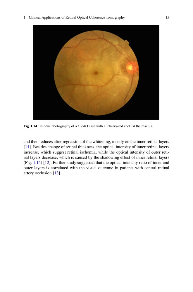
Fig. 1.14 Fundus photography of a CRAO case with a ‘cherry-red spot’ at the macula
and then reduces after regression of the whitening, mostly on the inner retinal layers
[11]. Besides change of retinal thickness, the optical intensity of inner retinal layers
increase, which suggest retinal ischemia, while the optical intensity of outer retinal layers decrease, which is caused by the shadowing effect of inner retinal layers
(Fig. 1.15) [12]. Further study suggested that the optical intensity ratio of inner and
outer layers is correlated with the visual outcome in patients with central retinal
artery occlusion [13].
15
Fig. 1.14 Fundus photography of a CRAO case with a ‘cherry-red spot’ at the macula
and then reduces after regression of the whitening, mostly on the inner retinal layers
[11]. Besides change of retinal thickness, the optical intensity of inner retinal layers
increase, which suggest retinal ischemia, while the optical intensity of outer retinal layers decrease, which is caused by the shadowing effect of inner retinal layers
(Fig. 1.15) [12]. Further study suggested that the optical intensity ratio of inner and
outer layers is correlated with the visual outcome in patients with central retinal
artery occlusion [13].