

11 Segmentation and Visualization of Drusen …
287
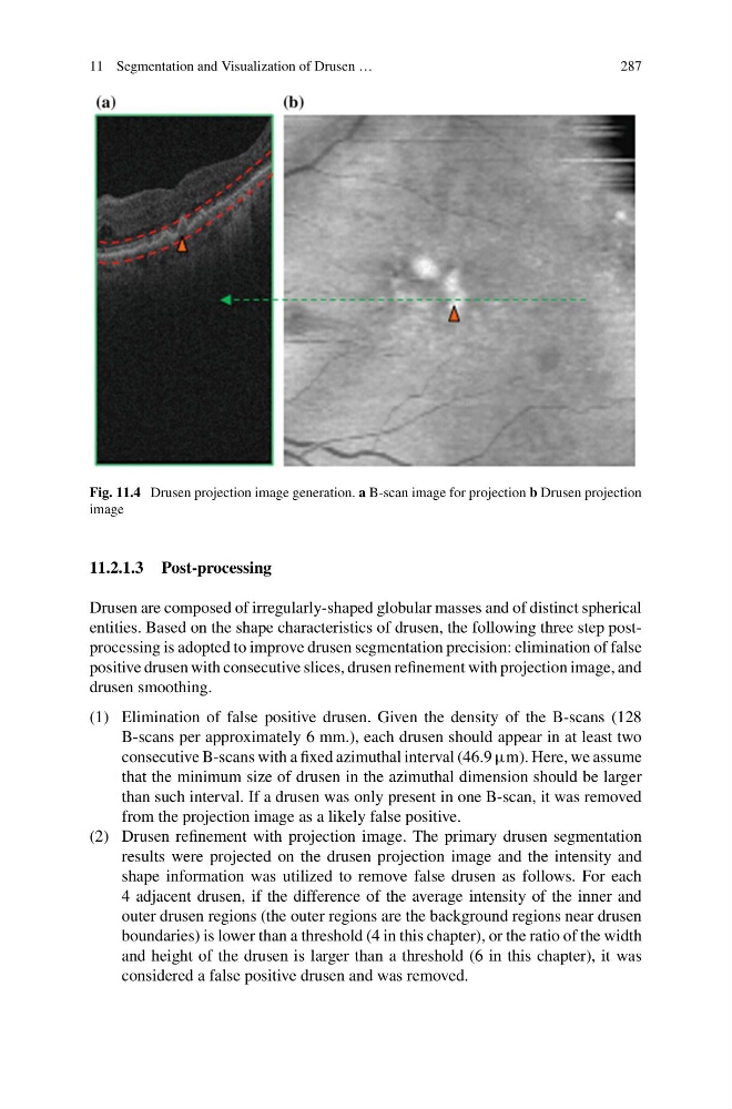
Fig. 11.4 Drusen projection image generation. a B-scan image for projection b Drusen projection
image
11.2.1.3 Post-processing
Drusen are composed of irregularly-shaped globular masses and of distinct spherical
entities. Based on the shape characteristics of drusen, the following three step postprocessing is adopted to improve drusen segmentation precision: elimination of false
positive drusen with consecutive slices, drusen refinement with projection image, and
drusen smoothing.
(1) Elimination of false positive drusen. Given the density of the B-scans (128
B-scans per approximately 6 mm.), each drusen should appear in at least two
consecutive B-scans with a fixed azimuthal interval (46.9 µm). Here, we assume
that the minimum size of drusen in the azimuthal dimension should be larger
than such interval. If a drusen was only present in one B-scan, it was removed
from the projection image as a likely false positive.
(2) Drusen refinement with projection image. The primary drusen segmentation
results were projected on the drusen projection image and the intensity and
shape information was utilized to remove false drusen as follows. For each
4 adjacent drusen, if the difference of the average intensity of the inner and
outer drusen regions (the outer regions are the background regions near drusen
boundaries) is lower than a threshold (4 in this chapter), or the ratio of the width
and height of the drusen is larger than a threshold (6 in this chapter), it was
considered a false positive drusen and was removed.
287
Fig. 11.4 Drusen projection image generation. a B-scan image for projection b Drusen projection
image
11.2.1.3 Post-processing
Drusen are composed of irregularly-shaped globular masses and of distinct spherical
entities. Based on the shape characteristics of drusen, the following three step postprocessing is adopted to improve drusen segmentation precision: elimination of false
positive drusen with consecutive slices, drusen refinement with projection image, and
drusen smoothing.
(1) Elimination of false positive drusen. Given the density of the B-scans (128
B-scans per approximately 6 mm.), each drusen should appear in at least two
consecutive B-scans with a fixed azimuthal interval (46.9 µm). Here, we assume
that the minimum size of drusen in the azimuthal dimension should be larger
than such interval. If a drusen was only present in one B-scan, it was removed
from the projection image as a likely false positive.
(2) Drusen refinement with projection image. The primary drusen segmentation
results were projected on the drusen projection image and the intensity and
shape information was utilized to remove false drusen as follows. For each
4 adjacent drusen, if the difference of the average intensity of the inner and
outer drusen regions (the outer regions are the background regions near drusen
boundaries) is lower than a threshold (4 in this chapter), or the ratio of the width
and height of the drusen is larger than a threshold (6 in this chapter), it was
considered a false positive drusen and was removed.