

264
F. Shi et al.
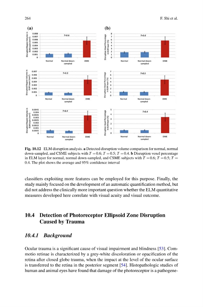
Fig. 10.12 ELM disruption analysis. a Detected disruption volume comparison for normal, normal
down-sampled, and CSME subjects with T 0.6; T 0.5; T 0.4. b Disruption voxel percentage
in ELM layer for normal, normal down-sampled, and CSME subjects with T 0.6; T 0.5; T
0.4. The plot shows the average and 95% confidence interval
classifiers exploiting more features can be employed for this purpose. Finally, the
study mainly focused on the development of an automatic quantification method, but
did not address the clinically more important question whether the ELM quantitative
measures developed here correlate with visual acuity and visual outcome.
10.4 Detection of Photoreceptor Ellipsoid Zone Disruption
Caused by Trauma
10.4.1 Background
Ocular trauma is a significant cause of visual impairment and blindness [53]. Commotio retinae is characterized by a grey-white discoloration or opacification of the
retina after closed globe trauma, when the impact at the level of the ocular surface
is transferred to the retina in the posterior segment [54]. Histopathologic studies of
human and animal eyes have found that damage of the photoreceptor is a pathogene-
F. Shi et al.
Fig. 10.12 ELM disruption analysis. a Detected disruption volume comparison for normal, normal
down-sampled, and CSME subjects with T 0.6; T 0.5; T 0.4. b Disruption voxel percentage
in ELM layer for normal, normal down-sampled, and CSME subjects with T 0.6; T 0.5; T
0.4. The plot shows the average and 95% confidence interval
classifiers exploiting more features can be employed for this purpose. Finally, the
study mainly focused on the development of an automatic quantification method, but
did not address the clinically more important question whether the ELM quantitative
measures developed here correlate with visual acuity and visual outcome.
10.4 Detection of Photoreceptor Ellipsoid Zone Disruption
Caused by Trauma
10.4.1 Background
Ocular trauma is a significant cause of visual impairment and blindness [53]. Commotio retinae is characterized by a grey-white discoloration or opacification of the
retina after closed globe trauma, when the impact at the level of the ocular surface
is transferred to the retina in the posterior segment [54]. Histopathologic studies of
human and animal eyes have found that damage of the photoreceptor is a pathogene-