

10 Layer Segmentation and Analysis for Retina with Diseases
265
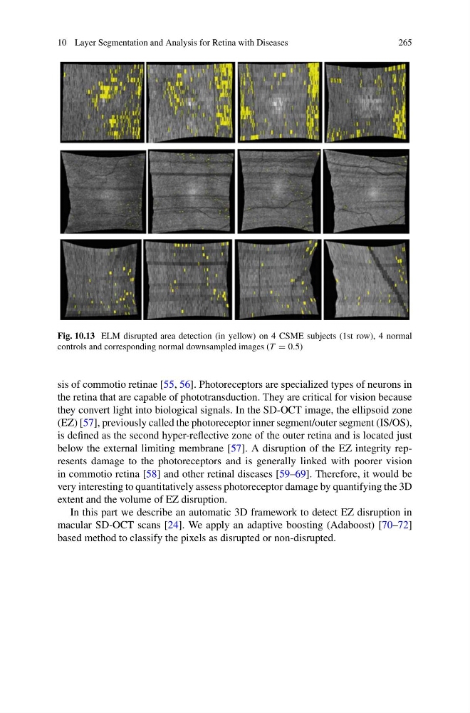
Fig. 10.13 ELM disrupted area detection (in yellow) on 4 CSME subjects (1st row), 4 normal
controls and corresponding normal downsampled images (T 0.5)
sis of commotio retinae [55, 56]. Photoreceptors are specialized types of neurons in
the retina that are capable of phototransduction. They are critical for vision because
they convert light into biological signals. In the SD-OCT image, the ellipsoid zone
(EZ) [57], previously called the photoreceptor inner segment/outer segment (IS/OS),
is defined as the second hyper-reflective zone of the outer retina and is located just
below the external limiting membrane [57]. A disruption of the EZ integrity represents damage to the photoreceptors and is generally linked with poorer vision
in commotio retina [58] and other retinal diseases [59–69]. Therefore, it would be
very interesting to quantitatively assess photoreceptor damage by quantifying the 3D
extent and the volume of EZ disruption.
In this part we describe an automatic 3D framework to detect EZ disruption in
macular SD-OCT scans [24]. We apply an adaptive boosting (Adaboost) [70–72]
based method to classify the pixels as disrupted or non-disrupted.
265
Fig. 10.13 ELM disrupted area detection (in yellow) on 4 CSME subjects (1st row), 4 normal
controls and corresponding normal downsampled images (T 0.5)
sis of commotio retinae [55, 56]. Photoreceptors are specialized types of neurons in
the retina that are capable of phototransduction. They are critical for vision because
they convert light into biological signals. In the SD-OCT image, the ellipsoid zone
(EZ) [57], previously called the photoreceptor inner segment/outer segment (IS/OS),
is defined as the second hyper-reflective zone of the outer retina and is located just
below the external limiting membrane [57]. A disruption of the EZ integrity represents damage to the photoreceptors and is generally linked with poorer vision
in commotio retina [58] and other retinal diseases [59–69]. Therefore, it would be
very interesting to quantitatively assess photoreceptor damage by quantifying the 3D
extent and the volume of EZ disruption.
In this part we describe an automatic 3D framework to detect EZ disruption in
macular SD-OCT scans [24]. We apply an adaptive boosting (Adaboost) [70–72]
based method to classify the pixels as disrupted or non-disrupted.