

248
F. Shi et al.
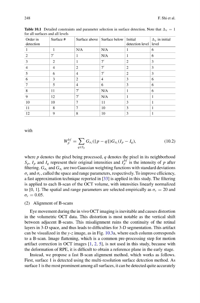
Table 10.1 Detailed constraints and parameter selection in surface detection. Note that x 1
for all surfaces and all levels
Order in
detection
Surface #
Surface above Surface below Initial
detection level
y in initial
level
1
1
N/A
N/A
1
6
2
7
1
N/A
1
6
3
2
1
7
2
3
4
4
2
7
2
3
5
6
4
7
2
3
6
3
2
4
3
6
7
5
4
6
3
6
8
11
7
N/A
1
6
9
12
7
N/A
1
1
10
10
7
11
3
1
11
8
7
10
3
1
12
9
8
10
3
1
with
W
b f
p
q∈S p
G σ s ( p − q)G σ r (I p − I q ),
(10.2)
where p denotes the pixel being processed, q denotes the pixel in its neighborhood
S p , I p and I q represent their original intensities and I
b f
p is the intensity of p after
filtering. G σ s and G σ r are two Gaussian weighting functions with standard deviations
σ s and σ r , called the space and range parameters, respectively. To improve efficiency,
a fast approximation technique reported in [33] is applied in this study. The filtering
is applied to each B-scan of the OCT volume, with intensities linearly normalized
to [0, 1]. The spatial and range parameters are selected empirically as σ s 20 and
σ r 0.05.
(2) Alignment of B-scans
Eye movement during the in vivo OCT imaging is inevitable and causes distortion
in the volumetric OCT data. This distortion is most notable as the vertical shift
between adjacent B-scans. This misalignment ruins the continuity of the retinal
layers in 3-D space, and thus leads to difficulties for 3-D segmentation. This artifact
can be visualized in the y-z image, as in Fig. 10.3a, where each column corresponds
to a B-scan. Image flattening, which is a common pre-processing step for motion
artifact correction in OCT images [1, 2, 5], is not used in this study, because with
the deformation of RPE, it is difficult to obtain a reference plane in the early stage.
Instead, we propose a fast B-scan alignment method, which works as follows.
First, surface 1 is detected using the multi-resolution surface detection method. As
surface 1 is the most prominent among all surfaces, it can be detected quite accurately
F. Shi et al.
Table 10.1 Detailed constraints and parameter selection in surface detection. Note that x 1
for all surfaces and all levels
Order in
detection
Surface #
Surface above Surface below Initial
detection level
y in initial
level
1
1
N/A
N/A
1
6
2
7
1
N/A
1
6
3
2
1
7
2
3
4
4
2
7
2
3
5
6
4
7
2
3
6
3
2
4
3
6
7
5
4
6
3
6
8
11
7
N/A
1
6
9
12
7
N/A
1
1
10
10
7
11
3
1
11
8
7
10
3
1
12
9
8
10
3
1
with
W
b f
p
q∈S p
G σ s ( p − q)G σ r (I p − I q ),
(10.2)
where p denotes the pixel being processed, q denotes the pixel in its neighborhood
S p , I p and I q represent their original intensities and I
b f
p is the intensity of p after
filtering. G σ s and G σ r are two Gaussian weighting functions with standard deviations
σ s and σ r , called the space and range parameters, respectively. To improve efficiency,
a fast approximation technique reported in [33] is applied in this study. The filtering
is applied to each B-scan of the OCT volume, with intensities linearly normalized
to [0, 1]. The spatial and range parameters are selected empirically as σ s 20 and
σ r 0.05.
(2) Alignment of B-scans
Eye movement during the in vivo OCT imaging is inevitable and causes distortion
in the volumetric OCT data. This distortion is most notable as the vertical shift
between adjacent B-scans. This misalignment ruins the continuity of the retinal
layers in 3-D space, and thus leads to difficulties for 3-D segmentation. This artifact
can be visualized in the y-z image, as in Fig. 10.3a, where each column corresponds
to a B-scan. Image flattening, which is a common pre-processing step for motion
artifact correction in OCT images [1, 2, 5], is not used in this study, because with
the deformation of RPE, it is difficult to obtain a reference plane in the early stage.
Instead, we propose a fast B-scan alignment method, which works as follows.
First, surface 1 is detected using the multi-resolution surface detection method. As
surface 1 is the most prominent among all surfaces, it can be detected quite accurately