

10
H. Chen et al.
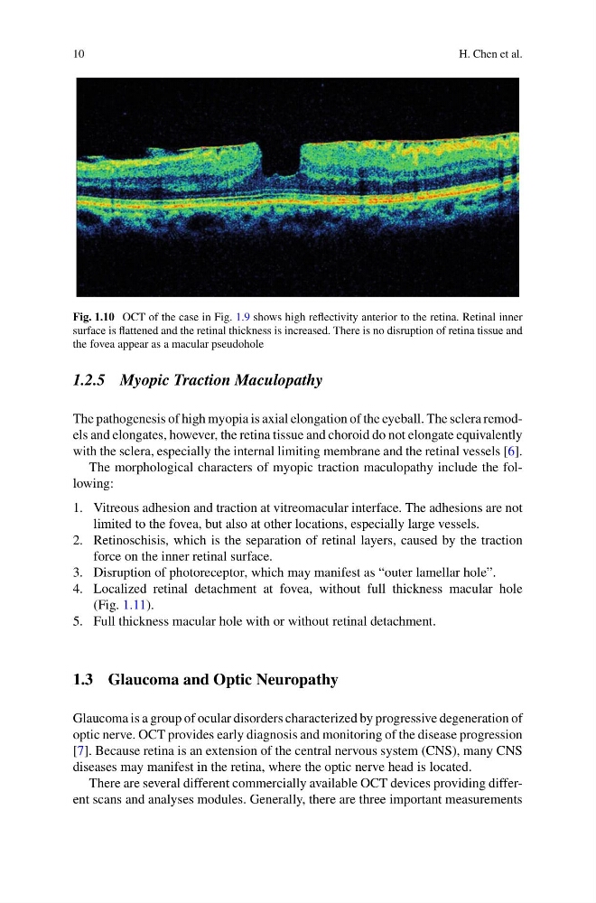
Fig. 1.10 OCT of the case in Fig. 1.9 shows high reflectivity anterior to the retina. Retinal inner
surface is flattened and the retinal thickness is increased. There is no disruption of retina tissue and
the fovea appear as a macular pseudohole
1.2.5 Myopic Traction Maculopathy
The pathogenesis of high myopia is axial elongation of the eyeball. The sclera remodels and elongates, however, the retina tissue and choroid do not elongate equivalently
with the sclera, especially the internal limiting membrane and the retinal vessels [6].
The morphological characters of myopic traction maculopathy include the following:
1. Vitreous adhesion and traction at vitreomacular interface. The adhesions are not
limited to the fovea, but also at other locations, especially large vessels.
2. Retinoschisis, which is the separation of retinal layers, caused by the traction
force on the inner retinal surface.
3. Disruption of photoreceptor, which may manifest as “outer lamellar hole”.
4. Localized retinal detachment at fovea, without full thickness macular hole
(Fig. 1.11).
5. Full thickness macular hole with or without retinal detachment.
1.3 Glaucoma and Optic Neuropathy
Glaucoma is a group of ocular disorders characterized by progressive degeneration of
optic nerve. OCT provides early diagnosis and monitoring of the disease progression
[7]. Because retina is an extension of the central nervous system (CNS), many CNS
diseases may manifest in the retina, where the optic nerve head is located.
There are several different commercially available OCT devices providing different scans and analyses modules. Generally, there are three important measurements
H. Chen et al.
Fig. 1.10 OCT of the case in Fig. 1.9 shows high reflectivity anterior to the retina. Retinal inner
surface is flattened and the retinal thickness is increased. There is no disruption of retina tissue and
the fovea appear as a macular pseudohole
1.2.5 Myopic Traction Maculopathy
The pathogenesis of high myopia is axial elongation of the eyeball. The sclera remodels and elongates, however, the retina tissue and choroid do not elongate equivalently
with the sclera, especially the internal limiting membrane and the retinal vessels [6].
The morphological characters of myopic traction maculopathy include the following:
1. Vitreous adhesion and traction at vitreomacular interface. The adhesions are not
limited to the fovea, but also at other locations, especially large vessels.
2. Retinoschisis, which is the separation of retinal layers, caused by the traction
force on the inner retinal surface.
3. Disruption of photoreceptor, which may manifest as “outer lamellar hole”.
4. Localized retinal detachment at fovea, without full thickness macular hole
(Fig. 1.11).
5. Full thickness macular hole with or without retinal detachment.
1.3 Glaucoma and Optic Neuropathy
Glaucoma is a group of ocular disorders characterized by progressive degeneration of
optic nerve. OCT provides early diagnosis and monitoring of the disease progression
[7]. Because retina is an extension of the central nervous system (CNS), many CNS
diseases may manifest in the retina, where the optic nerve head is located.
There are several different commercially available OCT devices providing different scans and analyses modules. Generally, there are three important measurements