

1 Clinical Applications of Retinal Optical Coherence Tomography
11
in retinal OCT for glaucoma and optic neuropathy, including parapapillary retinal
nerve fiber layer thickness, retinal ganglion cells complex layer thickness, and morphology of optic nerve head.
1.3.1 Parapapillary Retinal Nerve Fiber Layer Thickness
Retinal nerve fibers are the axons of retinal ganglion cells which make up the retinal
nerve fiber layer (RNFL) lying just below the internal limiting membrane. RNFL
has high reflectivity and can be easily identified and segmented on OCT images.
The nerve fibers exit the eyeball and connect to the brain via optic nerve. Therefore,
RNFL thickness is the highest at the parapapillary regions. Typically, OCT provides
a cube scan centered on the optic nerve head and produces a RNFL thickness map.
The RNFL thickness at the 3.4 mm diameter circle is calculated at each pixel. The
average thickness at each clock hour and quadrant are also calculated. The results
have been compared to the distribution of normal database and displayed as false
color scale. The symmetry of RNFL thickness has been calculated [8]. In early stage
of glaucoma, the RNFL reduced mostly on the superotemporal and inferotemporal
bundles, but the late stage, RNFL thinning involves the entire region (Fig. 1.12).
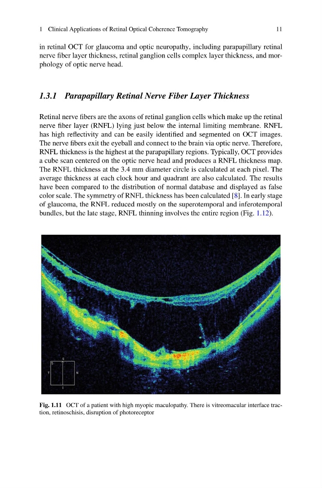
Fig. 1.11 OCT of a patient with high myopic maculopathy. There is vitreomacular interface traction, retinoschisis, disruption of photoreceptor
11
in retinal OCT for glaucoma and optic neuropathy, including parapapillary retinal
nerve fiber layer thickness, retinal ganglion cells complex layer thickness, and morphology of optic nerve head.
1.3.1 Parapapillary Retinal Nerve Fiber Layer Thickness
Retinal nerve fibers are the axons of retinal ganglion cells which make up the retinal
nerve fiber layer (RNFL) lying just below the internal limiting membrane. RNFL
has high reflectivity and can be easily identified and segmented on OCT images.
The nerve fibers exit the eyeball and connect to the brain via optic nerve. Therefore,
RNFL thickness is the highest at the parapapillary regions. Typically, OCT provides
a cube scan centered on the optic nerve head and produces a RNFL thickness map.
The RNFL thickness at the 3.4 mm diameter circle is calculated at each pixel. The
average thickness at each clock hour and quadrant are also calculated. The results
have been compared to the distribution of normal database and displayed as false
color scale. The symmetry of RNFL thickness has been calculated [8]. In early stage
of glaucoma, the RNFL reduced mostly on the superotemporal and inferotemporal
bundles, but the late stage, RNFL thinning involves the entire region (Fig. 1.12).
Fig. 1.11 OCT of a patient with high myopic maculopathy. There is vitreomacular interface traction, retinoschisis, disruption of photoreceptor