

1 Clinical Applications of Retinal Optical Coherence Tomography
9
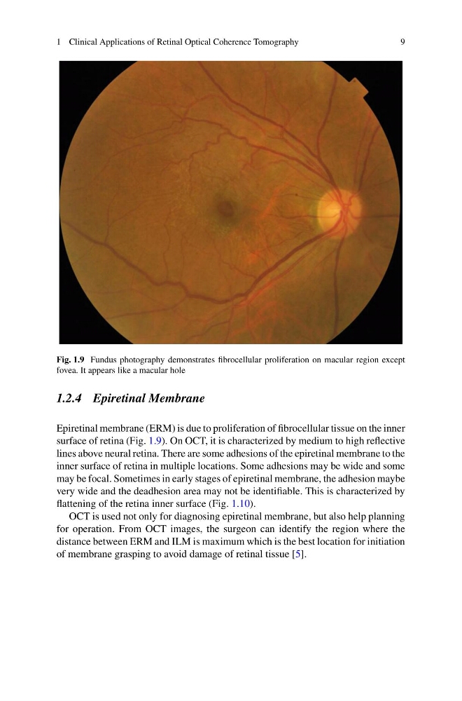
Fig. 1.9 Fundus photography demonstrates fibrocellular proliferation on macular region except
fovea. It appears like a macular hole
1.2.4 Epiretinal Membrane
Epiretinal membrane (ERM) is due to proliferation of fibrocellular tissue on the inner
surface of retina (Fig. 1.9). On OCT, it is characterized by medium to high reflective
lines above neural retina. There are some adhesions of the epiretinal membrane to the
inner surface of retina in multiple locations. Some adhesions may be wide and some
may be focal. Sometimes in early stages of epiretinal membrane, the adhesion maybe
very wide and the deadhesion area may not be identifiable. This is characterized by
flattening of the retina inner surface (Fig. 1.10).
OCT is used not only for diagnosing epiretinal membrane, but also help planning
for operation. From OCT images, the surgeon can identify the region where the
distance between ERM and ILM is maximum which is the best location for initiation
of membrane grasping to avoid damage of retinal tissue [5].
9
Fig. 1.9 Fundus photography demonstrates fibrocellular proliferation on macular region except
fovea. It appears like a macular hole
1.2.4 Epiretinal Membrane
Epiretinal membrane (ERM) is due to proliferation of fibrocellular tissue on the inner
surface of retina (Fig. 1.9). On OCT, it is characterized by medium to high reflective
lines above neural retina. There are some adhesions of the epiretinal membrane to the
inner surface of retina in multiple locations. Some adhesions may be wide and some
may be focal. Sometimes in early stages of epiretinal membrane, the adhesion maybe
very wide and the deadhesion area may not be identifiable. This is characterized by
flattening of the retina inner surface (Fig. 1.10).
OCT is used not only for diagnosing epiretinal membrane, but also help planning
for operation. From OCT images, the surgeon can identify the region where the
distance between ERM and ILM is maximum which is the best location for initiation
of membrane grasping to avoid damage of retinal tissue [5].